Home
| Quotations
| Misc
Notes | Notes
2 | Hair
| DemicDiff
| Diversity
| DNA|
Asian
IQ | Keita2008
data | Blood | Tropical
Civ
Egypt in Africa
| Black-Greek-DNA links
| Notes 3
|Notes 4| Notes
5 | Notes
6 | Notes 7 | Misc
news clips
|
Ethiopians
| Nubians
|
African
Tmeline
Misc news stories and clips
TIMES-ONLINE NEWS ARTICLE -
Skull of Cleopatra's sister
Murdered sister of Cleopatra found- a 2009 news
article reveals details about Arsinoe, the sister Cleopatra and
Mark Anthony had murdered as a potential rival.
QUOTE:
"Caroline
Wilkinson, a forensic anthropologist, reconstructed the missing
skull based on measurements taken in the 1920s. Using computer
technology it was possible to create a facial impression of what
Arsinöe might have looked like.
“It has got this long head shape,” said Wilkinson.
“That’s something you see quite frequently in ancient
Egyptians and black Africans. It could suggest a mixture of
ancestry.”
www.timesonline.co.uk/tol/news/world/middle_east/article5908494.ece
BBC NEWS ARTICLE- 2009 - skeleton of
Cleopatra's sister
In BBC news article, Austrian scientists say skeleton of Cleopatra's sister
was African.
QUOTE:
"Elizabeth Taylor's European Cleopatra
persists in the public imagination Cleopatra, the last Egyptian Pharaoh,
renowned for her beauty, was part African, says a BBC team which believes it has
found her sister's tomb.
Queen Cleopatra was a descendant of Ptolemy, the Macedonian general who ruled
Egypt after Alexander the Great. But remains of the queen's sister Princess
Arsinoe, found in Ephesus, Turkey, indicate that her mother had an
"African" skeleton.
Experts have described the results as "a real
sensation."
The discovery was made by Hilke Thuer of the Austrian
Academy of Sciences.
"It is unique in the life of an archaeologist to
find the tomb and the skeleton of a member of Ptolemaic dynasty," she
said.. "That Arsinoe had an African mother is a real sensation which
leads to a new insight on Cleopatra's family and the relationship of the sisters
Cleopatra and Arsinoe."
news.bbc.co.uk/2/hi/also_in_the_news/7945333.stm
Some DNA researchers claim an African origin for the Arabic
languages
Report:
archiver.rootsweb.ancestry.com/th/read/GENEALOGY-DNA/2002-05/1022778928
Near Eastern languages came from Africa 10,000 years ago
Investigator: Ene Metspalu
Tuesday May 28th, 2002
by Laura Spinney
Analysis of thousands of mitochondrial DNA samples has led Estonian
archeogeneticists to the origins of Arabic. Ene Metspalu of the
Department of Evolutionary Biology at Tartu University and the
Estonian Biocentre in Tartu, claims to have evidence that the Arab-
Berber languages of the Near and Middle East came out of East Africa
around 10,000 years ago. She has found evidence for what may have
been the last sizeable migration out of Africa before the slave
trade.
Genetic markers transmitted through either the maternal or paternal
line have been used to trace the great human migrations since Homo
sapiens emerged in Africa. But attempts to trace the evolution of
languages have met with less success, partly because of the impact on
languages of untraceable political and economic upheavals.
Metspalu and colleagues analyzed inherited variations in a huge
number of samples - almost 3000 - of mitochondrial DNA (mtDNA) taken
from natives of the Near East, Middle East and Central Asia, as well
as North and East Africa.
mtDNA is inherited through the maternal line, and by comparing their
data with existing data on European, Indian, Siberian and other
Central Asian populations, the researchers were able to create a
comprehensive phylogenetic map of maternal lineages diverging from
Africa and spreading towards Europe and Asia.
Working in collaboration with language specialists, they found that
this movement 10,000 years ago, which was probably centred on
Ethiopia, could well have been responsible for seeding the Afro-
Asiatic language from which all modern Arab-Berber languages are
descended.
"This language was spoken in Africa 10,000 or 12,000 years ago,"
Metspalu told BioMedNet News. "We think it was around that time that
carriers brought these Afro-Asiatic languages to the Near East." The
language, or its derivatives, later spread much further afield.
What could have triggered the movement she can only speculate. One
possibility is that increasing desertification was causing famine in
Africa and driving hunters further afield in search of animals.
Interestingly, the lineages they traced through this 10,000-year-old
migration didn't seem to get much further north than modern-day Syria
or east of modern-day Iraq. There is no evidence of the lineages in
the mtDNA of people from Turkey or Iran, says Metspalu.
"We can't understand why this boundary [to the Arab-Berber speaking
world] is so sharp," she said. "They came out of Africa, and when
they reached Turkey they just stopped." She believes some kind of
physical boundary, now vanished, must have impeded them.
The same genetic detective work has confirmed archeological evidence
that the biggest movement out of Africa occurred around 50,000 years
ago - which is when Africans first settled in other continents - and
that it originated in a small East African population.

First modern Europeans looked like Africans
www.telegraph.co.uk/news/worldnews/europe/romania/5273654/Scientists-reveal-face-of-the-first-European.html
Scientists reveal face of the first European
The face of the first European has been recreated from bone fragments by scientists.
By Urmee Khan, Digital and Media Correspondent
Published: 8:22PM BST 04 May 2009
The first modern European Forensic artist Richard Neave reconstructed the face based on skull fragments from 35000 years ago. Photo: BBC The head was rebuilt in clay based on an incomplete skull and jawbone discovered in a cave in the south west of the Carpathian Mountains in Romania by potholers.
Using radiocarbon analysis scientists say the man or woman, it is still not possible to determine the sex, lived between 34,000 and 36,000 years ago.
Europe was then occupied by both Neanderthal man, who had been in the region for thousands of years, and anatomically-modern humans Homo sapiens.
Modern humans first arrived in Europe from Africa.
The skull appears very like humans today, but it also displays more archaic traits, such as very large molar teeth, which led some scientists to speculate the skull may belong to a hybrid between Homo sapiens and Neanderthals an idea discounted by other experts.
Erik Trinkaus, professor of anthropology at Washington University in Missouri, said the jaw was the oldest, directly-dated modern human fossil. "Taken together, the material is the first that securely documents what modern humans looked like when they spread into Europe," he said.
The model was created by Richard Neave, a forensic artist, for a BBC programme about the origins of the human race and evolution.
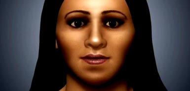
DISCOVERY CHANNEL Reconstruction of Queen
Nefertiti Forensic reconstruction team of British experts
did not know the ethnicity or origin of skull in advance - USA Today Article

|
|
|
|
Could this be the profile of a queen?
By Tim Friend, USA TODAY
Is this Nefertiti? Two months ago, a team of Egyptologists led by
British scientist Joann Fletcher of the University of York announced
that a neglected mummy collecting dust in a nondescript tomb was
actually that of ancient Egypt's most famous female ruler.
In an effort to confirm her identity, two British
experts have applied their forensic skills to digital X-rays of the skull.
(Related graphic: Reconstructing
Nefertiti)
Neither Damian Schofield of Nottingham University
nor Martin Evison of Sheffield University knew in advance the identity of
their "victim." They specialize in reconstructing human faces
from skulls for murder cases in which the victim is unknown.
Schofield and Evison created a 3-D computer mesh of
the skull, then placed a series of markers to designate where tissue would
be added. Next, they added facial muscles to give the face its full depth
and contour. Finally, a graphic artist added skin texture, eye color, lips
and the crown.
Schofield and Evison say the reconstruction does
not prove the skull belongs to Nefertiti. But they were surprised at the
similarities with Nefertiti's bust, which was made during her lifetime and
is displayed at the Egyptian Museum in Berlin.
Says Fletcher: "I was bowled over by it, to be
honest. The face is that of a very strong individual indeed. She has such
a beautiful profile. She is stunning."
Nefertiti's image is one of the most popular today
from ancient Egypt. But the real queen was hated by Egyptian society after
her reign ended. An unusually powerful queen, she reigned with her
husband, Akhenaten, who ruled from 1352 to 1336 B.C., during the late 18th
dynasty. Nefertiti may have ruled as pharaoh for three years after his
death.
Nefertiti vanished from Egyptian history with no
trace of a royal tomb or evidence of a burial.
|
Link to Discovery Channel
dsc.discovery.com/egypt/nefertiti-face/face.html
Italian reconstruction team
confirms that Nefertiti bust in German museum is
covered with a plaster facade. CAT scans revealed another image beneath the
first layer.
Italian team's forensic reconstruction is shown below.
| An Italian duo have revealed what they claim is the 'real' face of Queen
Nefertiti.
Ethnologist Franco Crevatin, from the University of Trieste, and cosmetics
expert Stefano Anselmo, started with a recent CAT scan of the famous
queen's bust, held in Berlin's newly-reopened
Neues Museum. The scan of 'Nonofret'
as she's known in Germany, appeared to show a second face, made of stone,
buried beneath the stucco top layer the world has come to adore. Using
computer imaging, Crevatin and Anselmo have made what they feel is a
faithful reproduction of the hidden face. And though differences are
subtle - shallower eye sockets, lines around the mouth and a tiny bump on
the bridge of the nose - the duo claim their version is closer to the real
Nefertiti.
'Reproducing the face of a queen who is surrounded by such
mystery required months of painstaking, detailed work,'' said Crevatin.
''It was particularly difficult given the number of entirely diverging
theories on her. Some even believe the bust in Berlin is a fake, while
others believe the queen only had one eye''. The bust of Nefertiti, who
was the chief wife of Egyptian Pharaoh Akhenaten, is the central
attraction at the newly reopened Neues Museum in Berlin. Thought to have
been created in 1,345 BC by the sculptor Thutmose, it was discovered in
the remains of his workshop on the banks of the Nile by a German
archaeological team in 1912. The Egyptologist Ludwig Borchardt brought it
to Germany the following year, although there are conflicting accounts of
whether he lied in order to do so. Egyptian authorities have suggested the
bust was smuggled out illegally and recently renewed a long-running
campaign for its return. But Germany insists it acquired the Egyptian
beauty lawfully and says it is too fragile to be sent back.
{Image: Neuves Museum, Berlin, Germany, 2009) www.lifeinitaly.com/node/12529
|
|
|
|
Image of the bust of Nefertiti on the left courtesy
the Neues Museum, Berlin, Germany. The image on the right is the reconstruction made by
Franco Crevatin and Stefano Anselmo.
|
CT IMAGES REVEAL THE HIDDEN FACE OF NEFERTITI
|
|
www.culturekiosque.com/art/news/nefertiti_bust_ct_images343.html
By Culturekiosque Staff
NEW YORK, 1 April 2009 - Using CT imaging to
study a priceless bust of Nefertiti, researchers have uncovered
a delicately carved face in the limestone inner core and gained
new insights into methods used to create the ancient masterpiece
and information pertinent to its conservation, according to a
study published in the April issue of Radiology .
"We acquired a lot of information on how the bust was
manufactured more than 3,300 years ago by the royal
sculptor," said the study's lead author Alexander Huppertz,
M.D., director of the Imaging Science Institute in Berlin,
Germany. "We learned that the sculpture has two slightly
different faces, and we derived from interpretation of the CT
images how to prevent damage of this extremely precious art
object."
Nefertiti, the wife of the Egyptian pharaoh Akhenaten, was
the most renowned Great Royal Wife of all 31 Egyptian dynasties.
Considered one of the greatest finds of ancient Egypt, the bust
of Nefertiti was discovered in 1912, during excavation of the
studio of famous royal sculptor Thutmose.
The Nefertiti bust consists of a limestone core covered in
layers of stucco of varying thickness. The bust was examined
using CT for the first time in 1992, but recent advances in CT
technology allowed the researchers to analyze the statue in 2007
with greater precision.
"CT has changed significantly since 1992," Dr.
Huppertz said. "We can now acquire three-dimensional (3-D)
images at a much higher resolution."
Dr. Huppertz and colleagues used a 64-section spiral CT
technique with submillimeter section thickness to examine the
bust and assess its conservation status, gain information on its
creation and provide a 3-D surface reformation of the inner
limestone sculpture.
The results showed that a multi-step process was used to
create the sculpture. The stucco layer on the face and ears is
very thin, but the rear part of the reconstructed crown contains
two thick stucco layers. CT images showed several fissures and
non-uniform bonding between the layers.
The inner limestone face was delicately sculpted and highly
symmetric. Compared to the outer stucco face, the inner face
exhibited some differences: less depth in the corners of the
eyelids, creases around the corner of the mouth and cheeks, less
prominent cheekbones and a slight bump on the ridge of the nose.
The ears on the inner sculpture were similar to those visible on
the exterior.
Thin-section CT was able to provide detailed images of the
inner structure in a completely nondestructive manner and showed
the limestone core to be not just a mold, but a skillfully
rendered work of art. Retouching the creases in the corners of
the mouth and smoothing the bump on the nose on the outer face
may have been the artist's choice and reflective of the
aesthetic ideals of that era.
CT findings also may be important in preventing future damage
to the bust. The findings of multiple, varying layers of stucco,
as well as fissures in the shoulders, lower surface of the bust
and rear of the crown, indicate vulnerable areas requiring very
careful handling, and pressure on the layers of thick stucco is
to be avoided.
"Noninvasive CT technology and very advanced 3-D
post-processing tools allow us greater insight into the internal
composition and conservation status of the sculpture," Dr.
Huppertz said. "This knowledge will greatly contribute to
the preservation of this priceless antiquity."
The Nefertiti bust is part of the collection of the Egyptian
Museum of Berlin and will be moved in October 2009 to the
recently restored New Museum in the historical center of Berlin.
Special Report in April 2009 issue of Radiology
Nondestructive
Insights into Composition of the Sculpture of Egyptian Queen
Nefertiti with CT. Collaborating with Dr.
Alexander Huppertz were Dietrich Wildung, Ph.D., Barry Kemp,
Tanja Nentwig, Patrick Asbach, M.D., Franz Maximilian Rasche,
M.D., and Bernd Hamm, M..D
Radiology. April 2009 251:233-240;
doi:10.1148/radiol.2511081175 |
|
Recent study finds that ancient Egyptians more
resembled Black Americans than White Americans, confirming similar older studies
on modern Egyptians. In either case, Black Americans were closer to Egyptians,
modern or ancient, than White Europeans or White Americans.
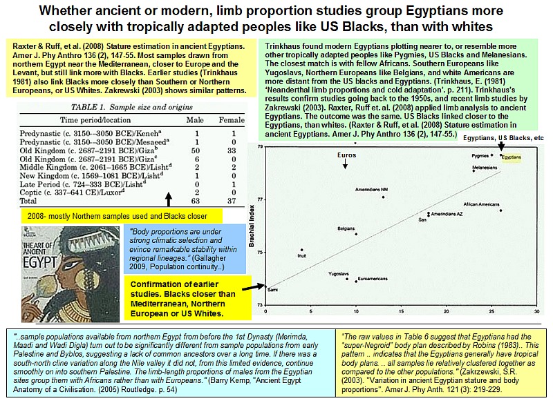
"Intralimb (crural and brachial) indices are significantly higher in ancient Egyptians than in American Whites (except crural index among females), i.e., Egyptians have relatively longer distal segments (Table 4). Intralimb indices are not significantly different between Egyptians and American Blacks... Many of those who have studied ancient Egyptians have commented on their characteristically tropical or African body plan (Warren, 1897;
Masali, 1972; Robins, 1983; Robins and Shute, 1983, 1984, 1986; Zakrzewski, 2003). Egyptians also fall within the range of modern African populations (Ruff and Walker, 1993), but close to the upper limit of modern Europeans as well, at least for the crural index (brachial
indices are definitely more African).. In terms of femoral and tibial length to total skeletal height proportions, we found that ancient Egyptians are significantly different from US Blacks, although still closer to Blacks than to Whites.
Comparisons of linear body proportions of Old Kingdom and non-Old Kingdom period individuals, and workers and high officials in our sample found no statistically significant differences among them. Zakrzewski (2003) also found little evidence for differences in linear body proportions of Egyptians over a wider temporal range. In general, recent studies of skeletal variation among ancient Egyptians support scenarios of biological continuity through time. Irish (2006) analyzed quantitative and qualitative dental traits of 996 Egyptians from Neolithic through Roman periods, reporting the presence of a few outliers but concluding that the dental samples appear to be largely homogeneous and that the affinities observed indicate overall biological uniformity and continuity from Predynastic through Dynastic and Postdynastic periods.
Zakrzewski (2007) provided a comprehensive summary of previous Egyptian craniometric studies and examined Egyptian crania from six time periods. She found that the earlier samples were relatively more homogeneous in comparison to the later groups. However, overall results indicated genetic continuity over the Egyptian Predynastic and Early Dynastic periods, albeit with a high level of genetic diversity within the population,
suggesting an indigenous process of state formation. She also concluded that while the biological patterning of the Egyptian population varied across time, no consistent temporal or spatial trends are apparent. Thus, the stature estimation formulae developed here may be broadly applicable to all ancient Egyptian populations.."
("Stature
estimation in ancient Egyptians: A new technique based on
anatomical reconstruction of stature." Michelle H. Raxter,
Christopher B. Ruff, Ayman Azab, Moushira Erfan, Muhammad Soliman, Aly El-Sawaf, (Am J Phys
Anthropol. 2008,
Jun;136(2):147-55
Recent study finds the ancient Egyptians had a
tropical body plan like sub-Saharan cold-adapted like European
type populations
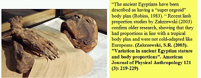
QUOTE:
"The raw values in Table 6 suggest
that Egyptians had the “super-Negroid” body plan
described by Robins (1983).. This pattern is supported by Figure
7 (a plot of population mean femoral and tibial lengths; data
from Ruff, 1994), which indicates that the Egyptians generally
have tropical body plans. Of the Egyptian samples, only the
Badarian and Early Dynastic period populations have shorter
tibiae than predicted from femoral length. Despite these
differences, all samples lie relatively clustered together as
compared to the other populations." (Zakrzewski,
S.R. (2003). "Variation in ancient Egyptian stature and body
proportions". American Journal of Physical Anthropology 121
(3): 219-229.
QUOTE(s):
 "Ancient Egyptian civilization was, in
ways and to an extent usually not recognized, fundamentally
African. The evidence of both language and culture reveals these
African roots. The origins of Egyptian ethnicity lay in the areas
south of Egypt. The ancient Egyptian language belonged to the
Afrasian family (also called Afroasiatic or, formerly,
Hamito-Semitic). The speakers of the earliest Afrasian languages,
according to recent studies, were a set of peoples whose lands
between 15,000 and 13,000 B.C. stretched from Nubia in the west
to far northern Somalia in the east. They
supported themselves by gathering wild grains. The first elements
of Egyptian culture were laid down two thousand years later,
between 12,000 and 10,000 B.C., when some of these Afrasian
communities expanded northward into Egypt, bringing with them a
language directly ancestral to ancient Egyptian. They also
introduced to Egypt the idea of using wild grains as food." (Christopher
Ehret (1996) "Ancient Egyptian as an African Language, Egypt
as an African Culture." In Egypt in Africa Egypt in Africa,
Theodore Celenko (ed), Indiana University Press)
"Ancient Egyptian civilization was, in
ways and to an extent usually not recognized, fundamentally
African. The evidence of both language and culture reveals these
African roots. The origins of Egyptian ethnicity lay in the areas
south of Egypt. The ancient Egyptian language belonged to the
Afrasian family (also called Afroasiatic or, formerly,
Hamito-Semitic). The speakers of the earliest Afrasian languages,
according to recent studies, were a set of peoples whose lands
between 15,000 and 13,000 B.C. stretched from Nubia in the west
to far northern Somalia in the east. They
supported themselves by gathering wild grains. The first elements
of Egyptian culture were laid down two thousand years later,
between 12,000 and 10,000 B.C., when some of these Afrasian
communities expanded northward into Egypt, bringing with them a
language directly ancestral to ancient Egyptian. They also
introduced to Egypt the idea of using wild grains as food." (Christopher
Ehret (1996) "Ancient Egyptian as an African Language, Egypt
as an African Culture." In Egypt in Africa Egypt in Africa,
Theodore Celenko (ed), Indiana University Press)
"Ancient Egypt belongs to a
language group known as 'Afroasiatic' (formerly called
Hamito-Semitic) and its closest relatives are other north-east
African languages from Somalia to Chad. Egypt's cultural
features, both material and ideological and particularly in the
earliest phases, show clear connections with that same broad
area. In sum, ancient Egypt was an African culture, developed by
African peoples, who had wide ranging contacts in north Africa
and western Asia." (Morkot, Robert (2005) The
Egyptians: An Introduction. Routledge. p. 10)
Levant vs the Horn
The Levant versus the Horn of Africa:
Evidence for Bidirectional Corridors of Human Migrations
J. R. Luis,1,2,* D. J. Rowold,1,* M. Regueiro,2 B. Caeiro,2 C. Cinnioğlu,3
C. Roseman,3 P. A. Underhill,3 L. L. Cavalli-Sforza,3 and R. J. Herrera1
Am J Hum Genet. 2004 March; 74(3): 532544
A more recent dispersal out of Africa, represented by the E3b-M35 chromosomes,
expanded northward during the Mesolithic (Underhill et al. 2001b). The East
African origin of this lineage is supported by the much larger variance of the
E3b-M35 males in Egypt versus Oman (0.5 versus 0.14; table 3). Consistent with
the NRY data is the mtDNA expansion estimate of 1020 ky ago for the East
African M1 clade. Local expansions of this clade and subsequent demic movements
may have resulted in the irregular presence of the M1 haplogroup in the
Mediterranean area (Quintana-Murci et al. 1999).
M35 chromosomes are seen in the Oman, North African, and East African
populations, as well as in the South African Khoisans (Underhill et al. 2000;
Cruciani et al. 2002; present study). There are three distinctive sublineages
(E3b1-M78, E3b2-M123, and E3b3-M81) that display nonrandom distributions (fig.
1). E3b1-M78 predominates in Egypt and Ethiopia, E3b3-M123 in Oman, and E3b2-M81
in northwestern Africa. Importantly, these three sublineages are restricted to
regions north of the equator. In contrast, the E3b*-M35 lineages appear to be
confined almost exclusively to the sub-Saharan populations, except for a very
low incidence in Egypt (2.7%) and a somewhat larger frequency in Ethiopia (7%,
as reported by Underhill et al. [2000]). The highest levels of E3b*-M35 are in
Tanzania (37.2%), Kenya (13.8%), and the Khoisans (11% in !Kung and 31% in Khwe).
The present-day Egyptian E3b-M35 distribution most likely results from a
juxtaposition of various demic episodes. Since the E3b*-M35 lineages appear to
be confined mostly to the sub-Saharan populations, it is conceivable that the
initial migrations toward North Africa from the south primarily involved
derivative E3b-M35 lineages. These include E3b1-M78, a haplogroup especially
common in Ethiopia (23%), and, perhaps, E3b2-M123 (2%), which is present as well
(Underhill et al. 2000; Cruciani et al. 2002; Semino et al. 2002). The data
suggest that two later expansions may have followed: one eastward along the
Levantine corridor into the Near East and the other toward northwestern Africa.
--------
"a Mesolithic population carrying Group III lineages with M35/M215
mutation [E3b] expanded northwards from sub-Saharan to north Africa
and the Levant" (Underhill et al., 2001, p. 55; see also Bosch et
al., 2001; Bar-Yosef, 1987) [Keita, 2005, p. 562]
The M35/M215 sub-clade cluster of haplotypes fragments a lineage (Ht
4) described previously (Hammer et al. 1997). We suggest that a
population with this sub-clade of the African YAP/M145/M203/PN2
cluster expanded into the southern and eastern Mediterranean at the
end of the Pleistocene...These lineages would have been introduced
then from the Middle East into southern Europe (and to a lesser
extent northern India and Pakistan) by farmers during the Neolithic
expansion. [Underhill et al., 2001, p. 51]
The Pleistocene epoch on the geologic timescale is the period from
1.8 million to 11,550 years BP [BP = before present (i.e. 1950)]
[en.wikipedia.org/wiki/Pleistocene]
...the diffusion of Neolithic farmers from the Near East between
4,000 and 7,500 years ago (Semino et al. 2000)...Interestingly, M35+
chromosomes (E3b*; or their evolutionary precursors E* and E3*) were
previously hypothesized to have migrated to Europe with farmers in
the Neolithic (Hammer et al. 1997; Rosser et al. 2000; Semino et al.
2000). However, because M35* chromosomes are rare in Europe, we
instead hypothesize that the derived lineage, E-M78 (E3b1), is the
more likely haplogroup reflecting Neolithic demic diffusion. [Behar
et al., 2003, p. 362]
In Europe E3b is the third largest group after "R" and "I"
haplogroups
Time-of-divergence estimates for E-M78δ chromosomes suggest a
relatively greater antiquity (14.7 ± 2.7 ky) for the separation of
eastern Africans from the other populations....demographic
population expansions involving clusters α [E-M78] in Europe
(TMRCA 7.8 ky; 95% CI 6.3-9.2 ky), β in northwestern Africa
(5.2 ky; 95% CI 3.2-7.5 ky), and γ in eastern Africa (9.6 ky;
95% CI 7.2-12.9 ky) should be considered the main contributors to
the relatively high frequency of haplogroup E-M78 in the surveyed
area. [Cruciani et al., 2004, pp. 1017-1018]
E3b originated in sub-Saharan Africa and expanded into the Near East
and northern Africa at the end of the Pleistocene (Underhill et al.
2001) E3b lineages would have then been introduced from the Near
East into southern Europe by immigrant farmers, during the Neolithic
expansion (Hammer et al. 1998; Semino et al. 2000; Underhill et al.,
2001). [Cruciani et al., 2004, pp. 1014-1015]
E3b's expansion into the Southern Levant may be connected to the
appearance of the Natufian Culture. [D'Agostino, 2006, p. 2]
...the clinal frequency distribution of E-M78α within Europe
testifies to important dispersal(s), most likely Neolithic or post-
Neolithic. These took place from the Balkans, where the highest
frequencies are observed, in all directions, as far as Iberia to the
west and, most likely, also to Turkey to the southeast. [Cruciani et
al., 2004, p. 1018]
E3b1-M78 is the most common haplogroup E lineage in Europe (Cruciani
et al. 2004; Semino et al. 2004). The spatial pattern...depicts a
nonuniform E3b1 geographic distribution with a frequency peak
centered in south Europe and SEE [South East Europe] (13%-16% in
southern Italians and 17%-27% in the Balkans) Declining frequencies
are evident toward western (10% in northern and central Italians),
central and eastern Europe (from 4% to 10% in Polish, Russians,
mainland Croatians, Ukrainians, Hungarians, Herzegovinians, and
Bosnians). Noteworthy is a low E3b1 frequency (5%) in Turkey.
Apart from its presence in Europe and the Middle East, E3b1 is also
found in eastern and northern Africa. Cruciani et al. (2004)
estimated that E3b-M78 might have originated in eastern Africa about
23.2 KYA (95% confidence interval [CI] 21.1-25.4)...Almost 93% of
SEE E3b1 chromosomes are clasified into α cluster. In Europe,
the highest E3b1α variance is among Apulians, Greeks, and
Macedonians, and the highest frequency of the cluster is among
Albanians, Macedonians, and Greeks...Furthermore, it may be
envisioned that the observed E3b1α frequency distribution in
Anatolia might stem from a back migration originating in south
Europe and SEE. Our estimated range expansion of 7.3 ± 2.8 KYA (95%
CI 6.3-9.2 KYA) estimate for expansions of cluster α
chromosomes in Europe reported by Cruciani et al. (2004) and the 6.4
KYA estimate for E3b1-M78 STR variance in Anatolia dated by
Cinnioğlu et al. (2004). The frequency and variance decline
of E3b1 in SEE is rather continuous..., with a frequency peak
extending from the southeastern edge of the region and a variance
peak in the southwest. Observed high E3b1 frequency in Kosovar
Albanians (46%) and Macedonian Romani (30%) represent a focal rather
than a clinal phenomenon resulting most likely from genetic drift.
E3b1 frequency and variance are significantly correlated with
latitude, showing higher values toward the south...A lower frequency
of E3b1 significantly distinguishes populations of the Adriatic-
Dinaric complex, i.e., mainland Croatians, Bosnians, and
Herzegovinians (7.9%; 95% CI 0.054-0.114), from their neighboring
populations of the Vardar-Morava-Danube river system, i.e., Serbians
and Macedonians (21.9%; 95% CI 0.166-0.283). These observations
hint a mosaic of different E3b1 dispersal modes over a short
geographic distance and point to the Vardar-Morava-Danube river
system as one of major routes for E3b1, in fact E3b1α,
expansion from south and southeastern to continental Europe. In
fact, dispersals of farmers throughout the Vardar-Morava-Danube
catchments basin are also evidenced in the archaeological record
(Tringham 2000). [Peričic et al., 2005]
Misc clips and articles - IQ data - Asian leadership
How DNA analysis undermines racial claims and
stereotypes

Scholars in the 1950s and 1960s such as Carleton Coons, claimed people like those depicted at left. the Tutsi of Rwanda in East Africa, were Caucasoid due to their height and narrow noses. Quote by Coons:
In Arabia prehistoric archaeology has barely been started. Yet we can be reasonably confident, until other evidence upsets the theory, that these deserts were the home of the slender variety of Caucasoid man. - Carleton Coons, The Story of man, 1954)
DNA analysis however undermines these and similar claims. Modern day genetic studies on the y-chromosome show the Tutsi to be fully indigenous African (80% e3a, 4% e3, 1% e3b and 15%B) with little to no East African genetic influence. The E3a DNA grouping is most prevalent in West Africa, not Europe, the Middle East or North Africa. Hence Africans do not need any race mix to have varying features. Indeed Africa is the most genetically diverse region on the planet (Tishkoff 2000). Quote:
To the west, Benin, Bamileke, and southern Cameroon are represented predominantly by chromosomes carrying the E3a-M2 mutation, a signature of the recent expansion of Bantu populations (Passarino et al. 1998; Scozzari et al. 1999; Underhill et al. 2001b; Cruciani et al. 2002). .. the E3a-M2 subclade is prevalent in our East African groups (Tutsi, Hutu, Kenya, and Tanzania) ..
(-- Luis, et al. (2004) The Levant versus the Horn. Am J Hum Genet. 2004; 74(3): 532544.)
Similar claims about Ethiopians are undermined by modern DNA. �The Encyclopedia Britannica (1962) article Ethiopia called them a white people with black Skin. DNA analysis by conservative mainstream scholars however undermines this claim, showing that while gene flow from Arabs and others occurred in some eras, the bulk of Ethiopian genes are [quote] predominant sub-Saharan African substrate. [Kivisild 2004 cites: (Cavalli-Sforza 1997; Passarino et al. 1998; Thomas et al. 2000; Cruciani et al. 2004; Luis et al. 2004). The overall genetic weight is thus with other sub-Saharan Africans.
2009 study finds Nubians the closest people ethnically to the ancient Egyptians
Quotes:
"The Mahalanobis D2 analysis uncovered close affinities between Nubians and Egyptians. Table 3 lists the Mahalanobis D2 distance matrix... In some cases, the statistics reveal that the Egyptian samples were more similar to Nubian samples than to other Egyptian samples (e.g. Gizeh and Hesa/Biga) and vice versa (e.g. Badari and Kerma, Naqada and Christian). .. The clustering of the Nubian and Egyptian samples together supports this paper's hypothesis and demonstrates that there may be a close relationship between the two populations. This relationship is consistent with Berry and Berry (1972), among others, who noted a similarity between Nubians and Egyptians.
Both mtDNA (Krings et al., 1999) and Y-Chromosome data (Hassan et al., 2008; Keita, 2005; Lucotte and Mercier, 2003) indicate that migrations, usually bidirectional, occurred along the Nile. Thus, the osteological material used in this analysis also supports the DNA evidence.
On this basis, many have postulated that the Badarians are relatives to South African populations (Morant, 1935 G. Morant, A study of predynastic Egyptian skulls from Badari based on measurements taken by Miss BN Stoessiger and Professor DE Derry, Biometrika 27 (1935), pp. 293-309.Morant, 1935; Mukherjee et al., 1955; Irish and Konigsberg, 2007). The archaeological evidence points to this relationship as well. (Hassan, 1986) and (Hassan, 1988) noted similarities between Badarian pottery and the Neolithic Khartoum type, indicating an archaeological affinity among Badarians and Africans from more southern regions. Furthermore, like the Badarians, Naqada has also been classified with other African groups, namely the Teita (Crichton, 1996; Keita, 1990).
Nutter (1958) noted affinities between the Badarian and Naqada samples, a feature that Strouhal (1971) attributed to their skulls possessing "Negroid" traits. Keita (1992), using craniometrics, discovered that the Badarian series is distinctly different from the later Egyptian series, a conclusion that is mostly confirmed here. In the current analysis, the Badari sample more closely clusters with the Naqada sample and the Kerma sample. However, it also groups with the later pooled sample from Dynasties XVIII-XXV.
Gene flow appears likely between the Egyptians and Nubians, although common adaptations to a similar environment may have also been a factor in their cranial similarities. This study does not rule out the possibility that in situ biological evolution occurred at other times not represented by the samples in this analysis. "
[end quotes]
-- Godde K. (2009) An Examination of Nubian and Egyptian biological distances: Support for biological diffusion or in situ development? Homo. 2009;60(5):389-404.
========================

www.newscientist.com/article/dn19094-tutankhamen-killed-by-sicklecell-disease.html?DCMP=OTC-rss&nsref=online-news
Tutankhamen 'killed by sickle-cell disease' claim scientists
King Tutankhamen, Egypt's boy king, was killed by the
inherited blood disorder sickle-cell disease not malaria. So says a
German team in what appears to be the best shot yet at solving the mystery
of the pharaoh's early demise.
From falling off a chariot to murder by poison, the
cause of Tutankhamen's
death has been a source of avid speculation since his mummified youthful
remains were discovered in 1922. He was 19 when he died around 1324 BC
after ruling for just nine years.
The first extensive scientific investigation of the
mummy was reported by Egypt's chief archaeologist Zahi Hawass and
colleagues earlier this year (JAMA,
vol 303, p 638). After running a battery of tests, including X-rays
and genetic analysis, they concluded that an inherited bone disorder
weakened the king, before an attack of malaria finished him off.
Key pieces of evidence were severe necrosis in the bones
of Tutankhamen's left foot, and the detection of genes from Plasmodium
falciparum, the parasite that causes malaria.
But in a
letter to JAMA this week, Christian
Timmann and Christian Meyer of the Bernhard Nocht Institute for
Tropical Medicine in Hamburg, Germany, suggest that Hawass's observations
can be explained much more elegantly by a diagnosis of sickle cell disease
(SCD).
Early death
People with SCD carry a mutation in the gene for
haemoglobin which causes their red blood cells to become rigid and
sickle-shaped. A single copy of the sickle-cell gene confers increased
immunity to malaria, so it tends to be common in areas where the infection
is endemic such as ancient Egypt. People with two copies of the gene
suffer severe anaemia and often die young.
Timmann and Meyer point out that SCD is the most common
cause of bone damage like Tutankhamen's. The deformed blood cells block
capillaries, preventing oxygen from reaching bone tissue. Tutankhamen's
parents are thought to be related, boosting the chance that they both
carried the sickle-cell gene.
People with SCD can still carry the malaria parasite in
their blood, despite their increased immunity. In King Tut's case, such an
infection could have triggered a fatal "sickle cell crisis" in
which his essential organs were starved of oxygen, Timmann says.
Members of Hawass's team describe the suggestion as
"interesting and plausible" and say that they are
"currently investigating". That would presumably require testing
Tutankhamen's mummy for the presence of the sickle-cell gene.
Timmann's lab has developed a test: given access to the
DNA, "we could do it in an hour", he says. But if that is the
line the Egyptians are taking, they are going it alone. Timmann says he
contacted them, offering to collaborate, but has received no reply.
Other researchers would also like to receive information
from the Egyptian team. In a second letter in JAMA this week, a
pair of US researchers suggests that King Tut and his relatives might
have had a hormonal disorder which causes, among other things,
deformed skulls and small male genitals. They need detailed photographs of
the skull of Tut's father to confirm their theory, but the Egyptian team
has exclusive access to the mummies and has not yet released the relevant
pictures.
Journal reference: JAMA,
vol 303, p 2473
Home
| Quotations
| Misc
Notes | Notes
2 | Hair
| DemicDiff
|
Egypt in Africa
| Black-Greek-DNA links