| Welcome
Definition
and Properties of Laser Light
How
a Laser Works
Laser
Types and Classifications
Laser Biological Hazards
Eyes
Skin
Non-Beam Laser Hazards
Protective Measures - Laser Safety
Protective Equipment
Path to Laser Operation
Test
Laser Links
|
|
Laser Biological Hazards
Eyes

Light causes biological damage through both temperature
effects due to absorbed energy and through photochemical reactions.
The chief mode of damage depends on the wavelength of the light and on the
tissue being exposed. For control of hazards from lasers, the damage
is believed to be due principally to temperature effects, and the critical
organs are the eye and the skin.
The Eye
The structure of the eye is shown below. The optical
components of the eye - those components that act together to focus an image
of an object on the retina - are the cornea, aqueous humor, lens, and
vitreous humor. The components of the eye most susceptible to laser
damage are the cornea, retina, and lens. The active components of the
eye are described in more detail below.
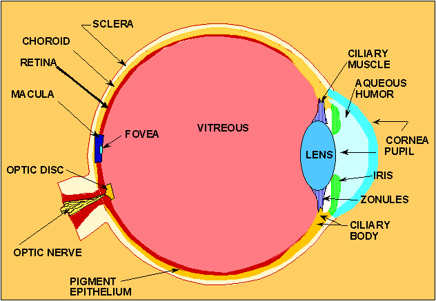
Components
-
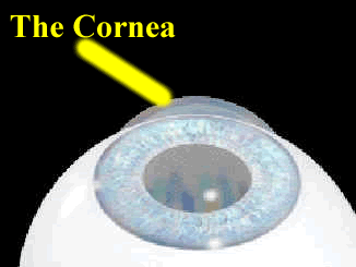
Cornea
-
Living tissue exposed directly to the
environmental elements. It is protected by a thin tear film.
-
The corneal epithelium has one of the
highest metabolic rates in the entire body. The tear layer of 6-10 um
thickness that protects this cell layer is fairly well balanced. The
outer most surface of the tear layer is a superficial lipid mono-multilayer
less than 0.5 um in thickness, then beneath this are mucin layers with
gradually increasing concentrations of mucin. The result is the cornea
has a mean index of refraction of 1.376. This provides
approximately 70% of the refractive power of the eye.
-
The cornea has a high metabolic
rate - rejuvenating itself in 24 to 48 hours.
-
Pupil - Iris - Sclera
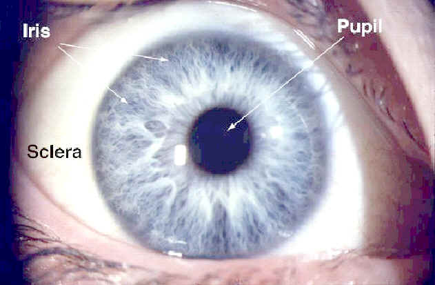
- Pupil
- Aperture of the eye.
- Normal range of 2 -
7 mm.
- Range decreases with age.
- 7 mm
is used for hazard calculations.
- Iris
- Adjusts the pupil of the eye.
- Circular, pigmented
membrane.
- Lies behind the
cornea.
- Sclera
- Dense fibrous shell.
- Maintains the roughly
spherical shape of the eye along with the internal pressure of
the eye.
-
Retina
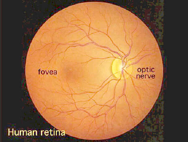
-
The retina is an
extension of the brain and consists of several complex layers of
nerve cells.
-
Made up of rods
and cones - rods for night and peripheral vision, cones for color
and resolution.
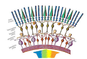
-
The macula is where the highest
resolution takes place. The cones have a yellowish pigment to
filter out blue light. Sharp vision is dependent on the
formation of a real image on the macula.
-
The fovea is in the center of the
macula and is where the cones are concentrated.
-
Lens
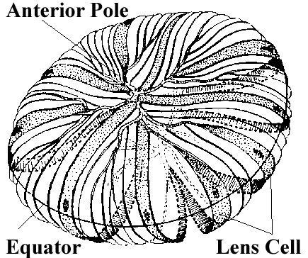
-
The crystalline
lens is supported in place by fine ligaments which are connected to
the ciliary body. The ciliary muscles control the eye's
focusing ability.
-
The lens is constructed of layers
of cells, similar to the make-up of an onion.
-
The lens provides fine tuning for
the eye. It provides approximately 30% of the refractive power
of the eye.
-
The lens has a slow
metabolism. Effects are delayed (cataracts). The lens
hardens and yellows with age.
Light Induced Biological Damage
Laser irradiation of the eye may cause damage to the
cornea, lens, or retina, depending on the wavelength of the light and the
energy absorption characteristics of the ocular tissues.
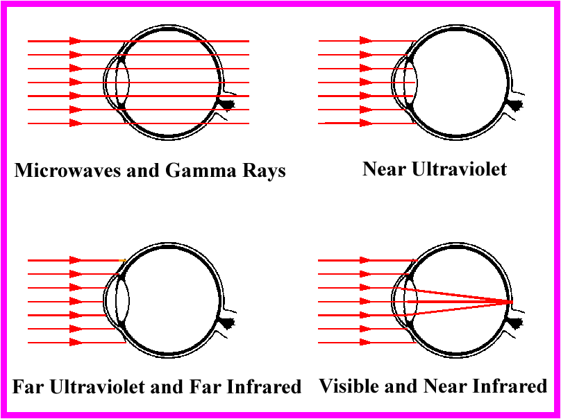
- The potential location of injury in the eye is
directly related to the wavelength of the laser radiation. For laser
radiation entering the eye:
- Near Ultraviolet Wavelengths (UVA) 315 - 400 nm
- Most of the radiation is absorbed in the
lens of the eye.
- The effects are delayed and do not occur for
many years (e.g.; cataracts).
- Far Ultraviolet (UVB) 280 - 315 nm and (UVC)
100 - 280 nm
- Most of the radiation is absorbed in the
cornea.
- Keratocojunctivitis (snow blindness/welder's
flash) will result if sufficiently high doses are absorbed.
- Visible (400 -760 nm) and Near Infrared
(760 - 1400 nm)
- Most of the radiation is transmitted to the
retina*.
- Overexposure may cause flash blindness or
retinal burns and lesions.
- Far Infrared (1400 nm - 1 mm)
- Most of the radiation is transmitted to the
cornea.
- Overexposure to these wavelengths will cause
corneal burns.
*NOTE:
Laser retinal injury can be severe because of the focal magnification
(optical gain) of the eye which is approximately 100,000 times. This means
that an irradiance of 1 mW/cm2 entering the eye will be effectively
increased to 100 W/cm2 when it reaches the retina.


|