 Our
Lady of
Our
Lady of
NCM101 –
Human Behavior
Introduction to Psychiatric Nursing
Mental illnesses are generally not caused by
ultra-structural or anatomical defect. Any anatomical defect in the brain and
the spinal cord can be referred as neurological disease and not a psychiatric
disease. Psychiatric diseases are mostly caused by neurobiological disturbances
in the brain that may cause interruption in man’s thinking.
Before we go to specific diseases, lets discuss first the basic
anatomy and physiology. The central nervous system is composed of the brain and
the spinal cord. The basic unit of the nervous system is the neuron.
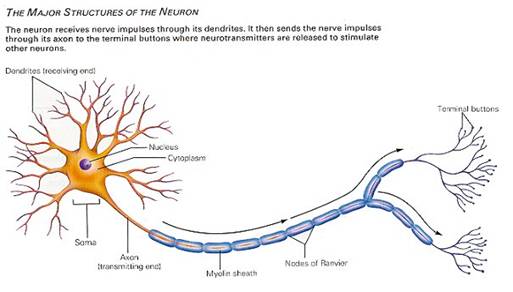
Many highly specialized types of neurons exist, and these
differ widely in appearance. Characteristically, neurons are highly asymmetric
in shape. Neurons consist of:
- The soma, or cell-body, the relatively large central
part of the cell between the dendrites and the axon.
- The axon, a much finer, cable-like projection which
may extend tens, hundreds, or even tens of thousands of times the diameter
of the soma in length. This is the structure which carries nerve signals
away from the neuron. Each neuron has only one axon, but this axon may
undergo extensive branching and thereby enable communication with many
target cells.
- The dendrite, a short, branching arbor of cellular
extensions. Each neuron has very many dendrites with profuse dendritic
branches. These structures form the main information receiving network for
the neuron.
Axon and dendrites alike are typically only about a
micrometer thick, while the soma is usually about 25 micrometers in diameter
and not much larger than the cell nucleus it contains. The axon of a human
neuron can be over a meter long, reaching from the base of the spine to the
toes.
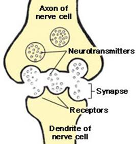
Neurons communicate with one another and to other cells
through synapses, where the axon tip of one cell impinges upon a dendrite or
soma of another, or less commonly to an axon.
Neurons communicate with one another across synapses.
This communication is usually chemically mediated by rapid secretion of
neurotransmitter molecules. Pre-synaptic neurons (i.e. the neurons which
release the neurotransmitter) may produce in the post-synaptic neurons (i.e.
the neurons being affected by the neurotransmitter) an electrical stimulation
(an electrical excitation) which will spread to the axon hillock
generating an action potential which then travels as a wave of
electrical excitation along the axon. Arrival of an action potential at the tip
of an axon triggers the release of neurotransmitter at a synaptic gap.
Neurotransmitters can either stimulate or suppress (inhibit) the electrical
excitability of a target cell. An action potential will only be triggered in
the target cell if neurotransmitter molecules acting on their post-synaptic
receptors cause the cell to reach its threshold potential.
The narrow cross-section of axons and dendrites lessens
the metabolic expense of carrying action potentials, although thicker axons convey
the impulses more rapidly, generally speaking.
Many neurons have insulating sheaths of myelin around
their axons, which enable their action potentials to travel faster than in
unmyelinated axons of the same diameter. Formed by glial cells in the central
nervous system and Schwann cells in the peripheral nervous system. The myelin
sheath in peripheral nerves normally runs along the axon in sections about
1 mm long, punctuated by unsheathed nodes of Ranvier.
Synapses are specialized junctions through which
cells of the nervous system signal to one another and to non-neuronal cells
such as muscles or glands.
Synapses form the circuits in which the neurons of the
central nervous system interconnect. They are thus crucial to the biological
computations that underlie perception and thought. They also provide the means
through which the nervous system connects to and controls the other systems of
the body.
Within the cells, small-molecule neurotransmitter
molecules are packaged in vesicles. When an action potential travels to the
synapse, the rapid depolarization causes calcium ion channels to open. Calcium
then stimulates the transport of vesicles to the synaptic membrane: the vesicle
and cell membrane fuse, leading to the release of the packaged neurotransmitter,
a mechanism called exocytosis.
A neurotransmitter's effect is determined by its
receptor. For example, GABA can act as a rapid or slow inhibitor, depending on
whether an ionotropic or metabotropic receptor is the target of the molecule. Small
molecule transmitters tend to have consistently inhibitory or excitatory action
on their targets. Meanwhile, the same polypeptide may have inhibitory or
excitatory effect on a cell, depending on the receptor.
Neurotransmitters may cause either excitatory or
inhibitory post-synaptic potentials. That is, they may help the initiation of a
nerve impulse in the receiving neuron, or they may discourage such an impulse,
by modifying the local membrane voltage potential. In the central nervous
system, combined input from several synapses is usually required to trigger an
action potential. Glutamate is the most prominent of excitatory transmitters;
GABA and glycine are well-known inhibitory neurotransmitters.
Many neurotransmitters are removed from the synaptic
cleft by a process is called reuptake (or often simply uptake).
Without reuptake, the molecules might continue to stimulate or inhibit the
firing of the postsynaptic neuron. Another mechanism for removal of a
neurotransmitter is digestion by an enzyme. For example, at cholinergic
synapses (where acetylcholine is the neurotransmitter) the enzyme
acetylcholinesterase breaks down the acetylcholine. Neuroactive peptides are
usually removed from the cleft by diffusion.
While some neurotransmitters (glutamate, GABA, glycine)
are used very generally throughout the central nervous system, others are only
used in certain brain regions by particular classes of nerve cells. Serotonin
is generally used as a neurotransmitter in cells involved in emotional
regulation. Dopamine acts as the neurotransmitter of choice for cells in the
hypothalamus which are effectively the brain's reward system, however it is
also involved in the control of movement.
Neurotransmitters which have these types of specific
actions are often targeted by drugs. Cocaine, for example, blocks the reuptake
of dopamine, leaving these neurotransmitters in the synaptic gap longer. Prozac
is a serotonin reuptake inhibitor, hence potentiating its effect. AMPT prevents
the conversion of tyrosine to L-DOPA, the precursor to dopamine; reserpine
prevents dopamine storage within vesicles; and deprenyl inhibits monoamine
oxidase (MAO) B and thus increases dopamine levels.
|
Neurotransmitter |
Abormal |
Disease Condition |
|
Dopamine |
increased |
Schizophrenia |
|
GABA |
Decreased |
Anxiety |
|
Serotonin |
decreased |
Depression |
Course Outline: prelims
| midterm
| finals
Handouts: week1 | week2 | week3
| week4 | week5
| week6 | week7
| week8 | week9 |
week10
| week11
| week12
| week 13 | week 14
Grades: Monday
| Tuesday
| Wednesday
| Thursday
| Friday
| Saturday