Multinodular disease: Too many nodules to easily count, with most of these nodules measuring < 1 cm in diameter.
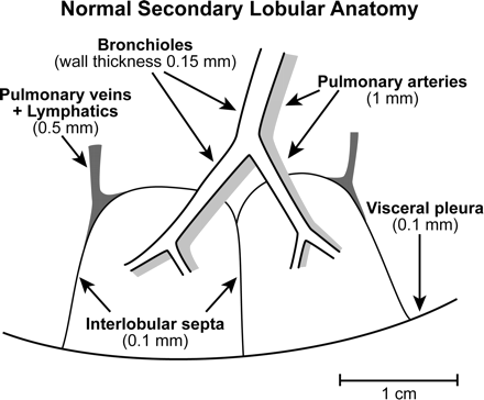
Secondary lobular anatomy. Secondary lobules represent fundamental anatomic units of the lung and are defined by centrilobular structures, including pulmonary arteries/arterioles and their accompanying bronchi/bronchioles, and peripheral structures, including the pulmonary veins and lymphatics within the interlobular septae.
For example, diseases such as sarcoidosis that localize within or adjacent to lymphatics predominate in those regions in which lymphatics are most extensive, specifically along the pleural and fissural surfaces, within the interlobular septae, and along the peribronchovascular axial interstitium.
Diseases that are primarily hematogenous in origin, such as miliary infections or hematogenous metastases, give rise to nodules that are randomly distributed throughout the secondary lobule, with the greatest profusion in the lung bases.
These patterns are clearly separate from nodules that result from inhalational disorders such as occur in patients with endobronchial spread of infection or hypersensitivity pneumonitis (HP), in which nodules are predominantly centrilobular in distribution.
******
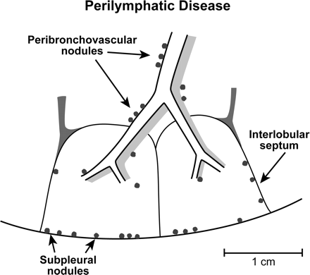
Perilymphatic disease. Nodules are preferentially subpleural, peribronchovascular within the axial interstitium, or along lobular septae. While this appearance is especially characteristic of nodular sarcoidosis, less commonly a similar pattern may also be seen in patients with silicosis or coal-workers pneumoconiosis.
******
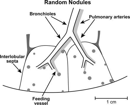
Random nodules. Lymphohematogenous disease. Random nodules are most commonly due to metastatic disease. Also, miliary infection.
Lymphangitic carcinomatosis, while hematogenous in origin, is easily distinguished from random metastatic nodules by the presence of characteristically thickened interlobular septae, preferentially involving the lung bases, and usually associated with asymmetric hilar adenopathy and pleural effusions.
******
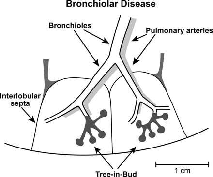
Bronchiolar disease. Bronchiolar inflammation resulting in so-called tree-in-bud opacities - dt extensive bronchiolar mucoid impaction.
Most importantly, unlike perilymphatic disease or random nodules, mucoid impacted bronchioles do not extend to the pleural, fissural, or septal surface. This pattern is nearly always due to infected secretions resulting from virtually any cause of acute or subacute bronchiolar infection.
******
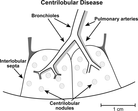
Centrilobular disease. Excluding those diseases that result in predominantly mucoid impaction due to infected secretions. The most common cause of diffuse centrilobular disease is subacute HP. This characteristically results in poorly defined, poorly marginated ground-glass opacities. Similar to tree-in-bud opacities, these rarely involve the pleural or fissural surfaces. While a number of different entities may result in predominantly centrilobular opacities, the differential diagnosis most often includes RB/RB-ILD. In distinction with subacute HP, RB in particular is less extensive, typically upper lobe in distribution, and almost always occurs in smokers.
This includes assessing a number of characteristics including whether nodules are as follows:
uniform or variable in size
sharply or poorly marginated
solid or subsolid in density (so-called ground-glass opacities)
or have a so-called tree-in-bud appearance
calcified, as occurs in fungal disease
or cavitary, e.g. septic emboli, metastatic disease, or Langerhans cell histiocytosis (LCH)
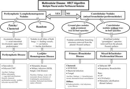
Step 1
Group 1:
Pleural or perifissural involvement characterize nodules as predominantly perilymphatic or lymphohematogenous in origin.
Step 2
Determining whether or not nodules are distributed diffusely or are patchy or clustered, with particular attention paid to the presence or absence of the extent of axial interstitial involvement.
Step 3
If nodules are clustered in a predominantly subpleural/axial distribution, they are deemed to be perilymphatic in distribution. The main disease to be considered is sarcoidosis. This diagnosis is further suggested by nodules that are typically ill-defined, frequently measuring only a few millimeters in size. Clusters of these nodules often have a "grainy" appearance and when sufficiently profuse may result in an appearance of poorly defined nodules or masses on corresponding chest radiographs (so-called alveolar sarcoid). When coalescent, these may simulate progressive massive fibrosis.
The most important differential diagnoses for this pattern of disease are silicosis and coal worker pneumoconiosis. In both of these occupational diseases, perilymphatic nodules are the primary abnormality, typically involving the mid and upper lung fields.
While lymphangitic carcinomatosis may result in perilymphatic nodules, in fact, CT scan findings are most often characterized by markedly thickened nodular interlobular septae usually asymmetrically involving the lower lobes and usually associated with adenopathy and effusions.
Step 4
If nodules prove to be diffuse instead of clustered, they are properly considered to be random in distribution.
hematogenous metastases. A basilar predominance is typically noted due to preferential blood flow to the lung bases. Individual nodules may have "feeding vessels" consistent with their hematogenous origin.
Nodules may also be either cavitary or surrounded by a "halo" of ground-glass attenuation, which is typical of hemorrhagic metastases such as those due to choriocarcinoma.
A number of malignancies can result in a miliary pattern, rendering differential diagnosis more problematic. This includes tumors, such as renal cell carcinoma, head and neck cancers, and testicular tumors, that have their primary venous drainage in the lungs.
The differential diagnosis includes a number of additional entities that result in random nodules. The most important of these is miliary infection.
Miliary metastases are frequently due to metastatic thyroid cancer, renal cancer, and melanoma, among other cancers, while larger less profuse metastases tend to be adenocarcinomas in adults, typically originating from the lung, breast, or the GI tract.
Less commonly, diffuse nodules may be identified in patients with septic emboli, invasive fungal infections, and pulmonary vasculitides. These entities frequently result in cavitary nodules, some with a distinct "halo" of ground-glass attenuation, and have even been described in patients with organizing pneumonia. Despite similarities between these entities and routine metastatic disease, it should be emphasized that the numbers of nodules identified in these cases usually fail to meet the criterion of "too many nodules to count," with the differential diagnosis again further aided by close clinical correlation.
Step 5
Group 2:
Anatomically, these nodules are centrilobular in distribution. Anatomically, these structures taper peripherally, stopping 5 to 10 mm short of the pleural or interlobular septal surfaces.
These nodules typically fall into the following two broad categories: "tree-in-bud" configuration and amorphous "ground-glass" nodules.
Step 6
If tree-in-bud configuration, infected mucoid impacted peripheral airways. Classically the result of the endobronchial spread of tuberculosis, this pattern may be seen in virtually any patient in whom there is infection of the peripheral airways. Not surprisingly, tree-in-bud opacities tend to be clustered rather than truly diffuse and frequently are associated with CT scan evidence of bronchiectasis.
Tree-in-bud opacities are nearly always the result of inspissated (ie, frequently aspirated) secretions lodged within centrilobular bronchioles, accounting for a branching configuration when coursing parallel to the CT scan plane.
Normal bronchioles, which have a diameter of < 1 mm and a wall thickness of < 0.1 mm, are below the limit of HRCT scan spatial resolution.
The presence of inspissated secretions results in bronchiolar distension and increased density, allowing their direct visualization. Another frequently encountered finding in patients with bronchiolar disease is so-called mosaic attenuation. In these cases, bronchiolar occlusion results in air-trapping, hypoxia of the poorly ventilated lung units with resultant reflex vasoconstriction. This combination of findings causes decreased attenuation of the affected areas of the lung with blood flow redistributed to normal lung.
While classically described in patients with an endobronchial spread of tuberculosis, in fact, tree-in-bud opacities can be identified in virtually any type of infectious bronchiolitis. This includes Mycobacterium tuberculosis, Mycobacterium avium-intracellulare, bacterial, viral, and fungal infections, and allergic bronchopulmonary mycosis. Differential diagnosis also includes follicular bronchiolitis, an entity that is characterized by the presence of hyperplastic lymphoid follicles and germinal centers occurring along the bronchovascular bundles.
Most often, infectious bronchiolitis results in clusters of tree-in-bud opacities. When they are widespread and diffuse, the differential diagnosis includes "Asiatic panbronchiolitis." This entity has a well-established predilection in Japanese, Chinese, and Korean populations, appears to show a genetic predisposition, and is usually seen in association with chronic sinusitis. Diffuse tree-in-bud opacities are also frequently encountered in patients with cystic fibrosis and viral bronchiolitis.
Step 7
Those cases in which centrilobular nodules are present in the absence of tree-in-bud opacities. Included in this category are a variety of diseases or "mixed" entities that have in common localization to the centrilobular portion of the secondary lobule. This includes diseases that primarily affect the centrilobular bronchiole, as well as those that are either primarily peribronchiolar or perivascular in origin.
Most often, this group of diseases results in a pattern of diffuse, poorly defined ground-glass nodules, which are typically the result of a primarily peribronchiolar distribution. The classic example of this appearance is subacute HP. This diagnosis is frequently first suggested on the basis of CT scan findings and is usually established by a combination of exposure history, clinical symptoms of a flu-like illness, the presence of specific serum antibodies when those data are available, increased numbers of lymphocytes and neutrophils in BAL fluid, and, when feasible, clinically significant improvement in symptoms when the patient is removed from the offending environmental agent.
The differential diagnosis encompasses a number of important disease entities, most importantly including RB, lymphocytic interstitial pneumonitis (LIP), and LCH. RB/RB-interstitial lung disease (ILD) are smoking-related disorders that may also result in poorly defined centrilobular nodules.
Also included in the differential diagnosis of individuals with diffuse centilobular nodules are diseases related to bronchiolar lymphatics. This includes mucosa-associated lymphoid tissue lymphoma (maltomas) and, in particular, LIP. LIP is most often seen in patients with underlying immunologic abnormalities, especially Sj�gren syndrome and AIDS, and is characterized histologically by diffuse hyperplasia of bronchus-associated lymphatic tissue, resulting in a diffuse, polyclonal lymphoid cell infiltrate surrounding the airways and expanding the lung interstitium. Poorly defined centrilobular nodules may also be seen early in the course of LCH. However, these most often are associated with characteristic bizarrely shaped, thick walled cysts, some of which represent cavitary nodules with characteristic sparing of the lung bases.
![]()
Approaches to Interpretation of Plain Radiographs
Approaches to Interpretation of CT
Approaches to Interpretation of MRI
Sample Normal Dictations
Sample Chest Dictations
Sample Nuclear Medicine Dictations
Normal Values
Chest Differentials
GI Differentials
Nuclear Medicine Gamuts
Chest Radiology Gamuts
Links
Multinodular Disease: A High-Resolution CT Scan Diagnostic Algorithm
![]()
![]()