 HOME
HOSPITAL
SERVICES & FACILITIES
COMMUNITY OUT-REACH
PATIENT GUIDE
PROCEDURES
HOME
HOSPITAL
SERVICES & FACILITIES
COMMUNITY OUT-REACH
PATIENT GUIDE
PROCEDURES
PUBLIC RELATIONS
EYE BANK
FEES & CHARGES
REPORTS
COURSES
LOCATION
CONTACT US
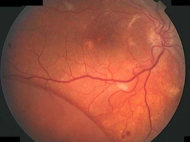
Retinal Detachment :
Normal Vision :
The retina is the nerve cell layer of the eye and acts much like film in a camera. When light enters the eye, it passes through the cornea and lens and is focused onto the retina. The retina transforms the light energy into vision and sends the information back to the brain through the optic nerve. The macula is the sensitive, central part of the retina and provides for sharp, detailed vision.
 Retinal Detachment :
In retinal detachment, the retina separates from the outer layers of the eye thus losing its function. If not treated early, retinal detachment may lead to impairment or complete loss of vision.
Cause :
Most retinal detachments are preceded by one or more tears or holes in the retina. Fluid passes through these openings and separates the retina from the adjacent layers of the eye. Near-sighted individuals are more commonly affected due to thinning of the retina. Holes or tears can then develop in the thinned retina. The vitreous (gel fluid in the eye) also plays a significant role by causing tugging on the retina especially when shrinkage occurs. Cataract surgery can also be precipitating cause. A positive family history of retinal detachment is another risk factor.
A combination of factors is usually responsible for retinal detachment.Retinal detachments can also be caused by other disease in the eye such as tumors, severe inflammations or complications of diabetes.
Symptoms :
Middle-aged and older persons may see floating black spots called floaters and flashes of light . In most cases, these symptoms do not indicate serious problems. In some eye, the sudden appearance of spots or flashes of light may herald the onset of retinal detachment. A thorough examination of the retina by an ophthalmologist after dilatation of the pupil is necessary to determine the cause of the symptoms.
Some retinal detachments can proceed unnoticed until a large section of the retina is detached. In these instances, patients may notice the appearance of a dark shadow in some parts of their vision. Further development of the retinal detachment will blur central vision and create and create significant sight loss in the affected eye.
A few detachments may occur suddenly and the patient will experience a total loss of vision in one eye. Similar rapid loss of vision may also be caused by bleeding into the vitreous, which may happen when the retina is torn.
Treatment :
If the retina is torn but detachment has not yet occurred, prompt treatment may prevent the occurrence of a complete detachment. Once the retina becomes detached, it must be repaired surgically by :
1) Laser photocoagulation
When new small retinal tears are found with little or no nearby retinal detachment, the tears are sometimes sealed with a laser light. The laser places small burns around the edge of the tear. These produce scars that seal the edges of the tear and prevent fluid from passing through and collecting under the retina.
2) Freezing or Cryopexy
Freezing through the sclera (white of the eye) behind a retinal tear will also stimulate scar formation and seal down the edges.
3) Surgical Repair
Successful reattachment of the retina consists of sealing the retinal tear with a silicone material, which is sutured to the sclera (white of the eye) to indent the eyeball inwards. Freezing applications are then used to bind the retina to the underlying layers.
Newer procedures have been developed to achieve the same result using the injection of a gas into the eye in suitable cases.
The surgery may be performed under local or general anaesthesia depending on the procedure, age and general health of the patient.
In more complex retinal detachments, it may be necessary to use a technique called Vitrectomy. This operation removes the vitreous body from the eye. In some cases, when the detached retina itself is severely shrunken or scarred, air or gas have to be used to fill the vitreous cavity temporarily.
Prognosis :
Over 90% of all retinal detachments can be reattached by modern surgical techniques. Occasionally, more than one operation may be required.The degree of vision which finally returns about six months after successful surgery depends upon a number of factors. In general, there is less visual return when the retina has been detached for a long time or if there is fibrous growth on the surface of the retina. Approximately 40% of successfully treated retinal detachments achieve excellent vision. The remainder attain varying amounts of reading vision. Due to continuous shrinkage of the vitreous and the development of fibrous growths on the retina, not all retinas can be attached. If the retina cannot be reattached, the eye will continue to lose sight and ultimately become blind.