Cu,Zn Superoxide Dismutase (SOD) is regularly a homodimeric enzyme,
whose Greek-key topology and structure are well preserved throughout
the evolutionary phyla.
Seven 3D structures of eukaryotic SODs have highlighted these concepts
and provided the structural bases for the study of the catalytic
mechanism, of substrate electrostatic guidance, of structural/thermal
stability and of the high affinity in subunit association.
In prokaryotic SODs this pattern is altered by amino acid
insertions/deletions and mutations which occur in loop regions,
altering the active site electrostatics and the subunit association
properties.
We have recently determined the 3D structure of E.coli
SOD at 2.0 Å resolution, and that of a new form of P.
leiognathi SOD at 2.1 Å
resolution. In agreement with solution studies, we found that E.coli SOD
provides the first known example of a SOD which is fully active in the
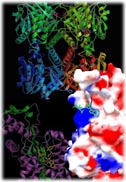
monomeric state. The region of molecular surface potentially involved
in the dimerization contacts is highly polar in E.Coli
SOD and displays a perturbed 3D structure, hampering the assembly of
the enzyme in a dimeric form. On the other hand, P.leiognathi
SOD, which also displays a polar patch on the molecular surface
involved in subunit association in the eukaryotic SODs, is found as a
dimeric enzyme. In this bacterial dimeric SOD, the association of
subunits is therefore based on an entirely new association surface
which buries several intervening water molecules.
Comparison of the different dimeric assemblies and of the monomeric
structure of E.coli SOD,
allows to underscore some general principles for the funcionality of an
enzyme which, in the framework of a reasonably conserved protein fold
topology, adopts different assembly rules in organisms which are not
necessarily distant along the evolutionary scale.
Engineering studies on P.leiognathi
SOD mutants at the subunit interface are in progress.
|