The Orbicularis Oculi
Understanding
the linchpin of myopia
More-click-polaris
Lock-of-dye? Poor-stick-of-Larry's Shock-of-pie?
Snore-flick-in-Paris Clock-you-fly?
I-drive-a-Lexus Ask-me-why?
No, no, no and no.
We're talking about the Orbicularis Oculi (say or-BICK-ye-Lair-iss
OCK-ye-li), one of the many muscles within
our face. This muscle is sometimes called the
Orbicularis Palpebrarum or the Sphincter Oculi.
What, Why
and Where?
The Orbicularis Oculi is a broad, thin, circular
muscle that covers the eyelid and the surrounding
eye socket. Because of its round shape, it's
considered a sphincter muscle. As you might
already know, sphincter muscles are ring-shaped
muscles that surround a natural opening in the
body. Their shape and function help to close the
opening that they cover.
The Orbicularis
Oculi is composed of three parts: the palpebral
portion, the orbital portion, and the lachrimal
portion. The palpebral portion is the part that
covers the eyelid. The orbital portion of the
muscle surrounds the orbit or eye socket,
extending from the bottom of the forehead down to
the front of the cheek. This muscle actually
connects to part of the upper jaw or maxilla. The
upper fibers of the orbital portion blend in with
the occipito-frontalis (on the forehead) and
corrugator supercilli (above the eyebrow)
muscles. The lachrimal portion (tensor tarsi;
Horner's muscle; Duverney's muscle) connects from
the inside bridge of the nose and passes across
the lachrimal sac to insert with the palpebral
portion of the muscle. These three portions work
together to narrow, close, or blink the eye.
X-Ray
Vision
The human face is one
massive maze of muscles. All facial muscles are
connected to the skin -- if this weren't the
case, then we would have a hard time making any
facial expressions. The reason why we're able to
express ourselves so easily with our face is
because of this amazing maze of muscular mayhem!
In order to help
you see what the Orbicularis Oculi muscle
actually looks like, I've enabled this webpage
with a high-tech, state-of-the-art, advanced,
bi-directional, titanium-plated, dual-action,
binary scanning, gamma-ray absorbing, x-ray
emitting mouse pointer that creates an x-ray
image when moved over a picture. Just 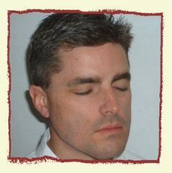 by coincidence, I happen to have a
picture of myself for you to test this out on. If
you move your technically advanced x-ray mouse
pointer over the picture, you will see the actual
muscles in my face. That big, round muscle that
goes around my eye is the Orbicularis Oculi.
Click on the picture if you'd like to see it
enlarged. by coincidence, I happen to have a
picture of myself for you to test this out on. If
you move your technically advanced x-ray mouse
pointer over the picture, you will see the actual
muscles in my face. That big, round muscle that
goes around my eye is the Orbicularis Oculi.
Click on the picture if you'd like to see it
enlarged.
The human body is
composed of about 650 muscles. In most cases,
muscles work in pairs. Consider the leg, for
example. One group of muscles work together to
draw the leg forward as you walk across the floor
or kick a soccer ball toward the goal. Another
group of muscles work together to draw the leg
backward while you walk or prepare to kick the
soccer ball again. This coordinated pairing of
muscle groups is found throughout our bodies.
This is what makes us "bendable" in
many different directions.
When one pair of
muscles move the body in a certain direction, the
antagonist pair of muscles that move the body in
the opposite direction must relax or else
conflict of movement will occur. This "I'll
relax to let you move" principle of yield is
known as Sherrington's Law of Reciprocal
Innervation. It was named after Sir Charles Scott
Sherrington, a British physiologist who shared
the Nobel Prize in 1932 for his studies into our
nervous and muscular systems. Therefore, when
contraction of a muscle is stimulated, there is a
simultaneous inhibition of its antagonist. This
is very essential for coordinated movement.
Sphincter muscles
are a little bit different, however. With a round
sphincter muscle such as the Orbicularis Oculi,
there is no direct antagonist to regulate its
movement. As pointed out by a Feldenkrais
practitioner friend of mine in Switzerland,
Dieter Didier Stoecklin, the relaxation of a
sphincter muscle is not governed by the
sensory-motor nervous system but by the
vegetative nervous system with its two branches
of sympathetic (arousal) and parasympathetic
(relaxing) branches. In other words, when the
Orbicularis Oculi muscle is pulled, there is no
opposing muscle to counteract the force and say,
"Hey, don't tread on me!"
Using your x-ray
imaging mouse cursor again, you'll notice that
there is a big muscle that goes all the way
around the mouth -- this muscle is called the
Orbicularis Oris ("orbicularis" because
it's round). You'll also notice that there are
two muscles on the cheek that connect the
Orbicularis Oculi muscle directly to the mouth.
The first of these is called the Levator Labii
Superioris (say le-VAY-ter LAB-ee-eye
soo-PEER-ee-or-iss) which is kind of
rectangular in shape. It connects near the lower
edge of the occular orbit (eye socket) and
attaches to the muscular tissue of the
orbicularis oris muscle of the upper lip. This
muscle is used to elevate the upper lip.
The second muscle
that connects the Orbicularis Oculi to your mouth
is called the Levator Labii Superioris Alaeque
Nasi (say le-VAY-ter LAB-ee-eye
soo-PEER-ee-or-iss uh-LEE-qwee NAYZ-eye), and
this is a bit more triangular in shape. It also
runs a bit along the side of the nose, hence the
"nasi" part of the name. This muscle is
used to draw the upper lip and nose upward, and
if you're ever expressing disdain or contempt,
you're using this muscle.
Why is it that
women often open their mouths while applying
make-up? Because they know that the mouth is
connected to the eye. If there is tension in the
mouth, then the levator muscles which connect the
mouth and the eye are shortened. When this
happens, the Orbicularis Oculi is pulled downward
which ultimately affects the shape of the eye.
"But how does
the Orbicularis Oculi affect eyeball shape?"
you ask. The Orbicularis Oculi circumscribes or
encircles the front half of the eyeball. The
eyeball protrudes from its socket all the through
the center of the Orbicularis Oculi. Take a look
at someone's profile, and you'll see how much the
eyeball protrudes through this muscle, almost as
if the eyeball had been "inserted" into
it. Slowly, over time, because of muscle tension,
this and other muscles in your body can change
shape because of tension. The tension does not
originate in the Orbicularis Oculi, but it
certainly ends up there.
And as stated
elsewhere, by reshaping the body through
relaxation, the shape of eyeball can be changed
and the vision improved.
As usual, stay
tuned for more.
DISCLAIMER: The
information presented on this website is for
informational purposes only.
|