Biology
Simulating X-Ray Diffraction Patterns of DNA Structure
Last update: 02/22/09 11:06 pm
 DNA
structure was solved by analyzing x-ray diffraction patterns of DNA crystals.
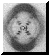
One of the crucial arguments in favor of
double helical structure was "Photo 51" which is shown on the left. Is it
possible to obtain an image similar to that of Photo 51 by computational
techniques, as a product of simulation?
DNA
structure was solved by analyzing x-ray diffraction patterns of DNA crystals.
One of the crucial arguments in favor of
double helical structure was "Photo 51" which is shown on the left. Is it
possible to obtain an image similar to that of Photo 51 by computational
techniques, as a product of simulation?
A naive representation of an x-ray
diffraction pattern is a
Fourier transform of molecular image. However, molecule is a 3D
entity that can be observed from various points in space. Thus the obvious
question is: what viewpoint on DNA crystal would produce an image similar
to that of "Photo 51" under Fourier transform? Although it is rather common to
find artistic renderings of DNA structure, they are not very useful in trying to
reproduce an x-ray diffraction pattern of the molecule. However, using the
calculations on double helical structure,
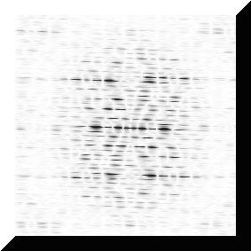 it
is possible to derive an abstract representation of DNA useful for Fourier
transform.
it
is possible to derive an abstract representation of DNA useful for Fourier
transform.
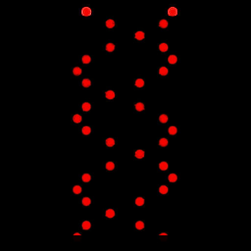
The right-side image represents red channel of the original DNA double helix. For simplicity, only the backbone of DNA was taken into consideration. Best results were achieved using the ball-and-stick model instead of space-filling model. The proposed segment was multiplied to obtain a longer version of the fiber. 2D Fourier transform was used to generate two projections (real and complex) which were combined to produce image on the left. The distinctive feature of "Photo 51" is clearly discernable.