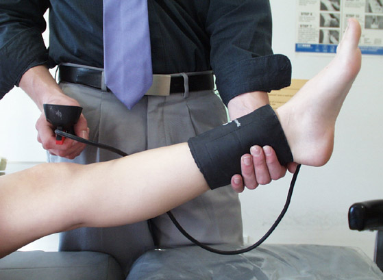
This test is done by
measuring blood pressure at the ankle and in the arm while a person is at
rest. Measurements are then repeated at both sites after 5 minutes of
walking on a treadmill.
By dividing the highest blood pressure at the ankle by the highest
recorded pressure in either arm, the ankle-brachial index (ABI) can be
calculated. The ABI result is used to predict the severity of peripheral
arterial disease (PAD) that may be present. A decrease in the ABI result
with exercise is a sensitive indicator that significant PAD is probably
present.
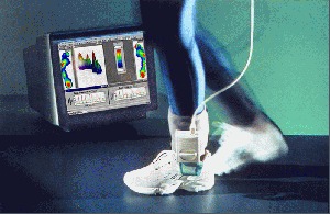
The analysis of dynamic or static pressure distribution revolves around Tekscan's innovative, real-time, tactile sensing systems. These systems are capable of measuring critical patient/surface interfaces with minimal interference. Extremely thin, flexible sensors accommodate most contours and provide highly accurate local pressure readings. Tekscan's pressure sensors provide the most appropriate spatial resolution required for the medical application under consideration. Vivid graphics, displayed on a PC, make the information easy to interpret and provide the clinician with excellent documentation.
- Achilles tendon/tendonitis/rupture/insertion
- Retrocalcaneal bursitis
- Plantar Fasciities/partial tear/chronic v. acute/lack of pathology for work comp/bilateral comparison
- Plantar Fibroma/with direct measurement
- Heel spur/fracture of spur/position on medial or lateral tuberosity
- Morton's neuroma/real time Mulder's click/differentiation from adjacent metatarsal bursitis
- Tibialis posterior/rupture/tendonitis/dysfunction
- Rheumatoid arthritis/capsular erosion/nodules
- Bone/near surface only/immediate view of stress fracture/chevron osteotomy/periostitis,subperiosteal hematoma, osteotomy bridging/motion at fx/osteotomy site/fx sesamoids,sesamoidal facet arthropathy
- Ankle sprain/deltoid ligament/partial vs. complete tears a.t.f./c.f./p.t.f.
- TCystic masses/fluid v. solid/compressibility/adjacent anatomic structure
- Fat pad thickness/diabetic at risk foot evaluation
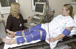
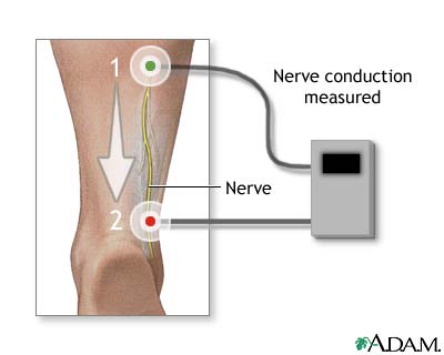
The NCV test can be used to detect true nerve disorders (such as neuropathy) or conditions whereby muscles are affected by nerve injury (such as carpal tunnel syndrome). Normal body temperature must be maintained for the NCV test, because low body temperatures slow nerve conduction
Current
Perception Threshold Test is a sensory test, which differentiates and
quantifies your pain. The test is painless but time consuming. The test
helps to diagnose cause of your pain and it helps to monitor the effects of
your treatment.
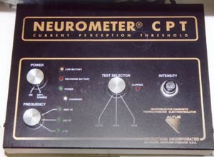
The
Neurometer utilizes three neuro-selective electrical stimuli to perform
extremely sensitive quantitative assessment sensory nerve fibers at any
cutaneous site.
The
current perception threshold (CPT) value is the minimum amount of a
current trans- cuatneously applied neuroselective current that a person
consistently perceives as evoking a sensation.
CPT
values are obtained for large diameter myelinated, small diameter myelinated
and unmyelinated sensory nerve fibers by stimulating with three different
neuroselectvie sine wave stimuli at 200Hz, 250Hz, and 5Hz, respectively. (Hz
=3D cycle per second)