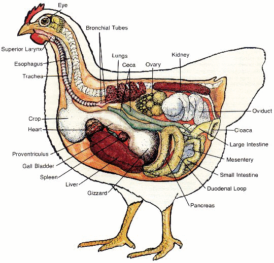
Respiratory System
Click To Enlarge
 Each nasal opening leads into a nasal cavity that is connected to sinus cavities around each eye. A split in the roof of the mouth provides an air passage between the nasal cavities and the lower respiratory system. The nasal cavities filter the air before it enters the lungs.
The larynx is located at the rear of the mouth. It is the structure connecting the trachea (windpipe) and gullet. The trachea is a tube that separates into two bronchial tubes, with each tube attached to a lung. The trachea and bronchial tubes are supported by rings of cartilage that prevent the tubes from collapsing.
The lungs are located near the vertebra and lay closely against the ribs. They resemble bright red sponges because of the abundant blood supply. Bird lungs are smaller in proportion to body size than other animals. Though small, the lungs are aided by an extensive system of air sacs found only in birds.
The air sacs are thin membrane sacs that surround the internal organs. They are used as reserve air space to increase lung capacity. When the bird's body is opened, the air sacs appear as clear thin membranes among the body organs. They are among the first sites affected by respiratory diseases.
Digestive System
The mouth is connected to the rest of the digestive system by a thin-walled tube called the esophagus or gullet. The lower portion of the esophagus forms a pouch called the crop. It functions as a temporary storage site for food. The lower end of the esophagus is attached to the bird's stomach.
The bird stomach has two parts -- proventriculus and gizzard. The proventriculus is the slightly enlarged area between the esophagus and gizzard. When opened it has a deeply textured appearance. The gizzard has a tough membrane inner lining firmly attached to the muscular outer part.
The lower end of the gizzard is attached to the upper end of the small intestine. The first portion of the small intestine is the duodenum. It is held in a loop-like position by the pancreas. The pink pancreas is located between and attached to the portions of the intestine forming the loop.
The lower portion of the small intestine is attached to a membrane called the mesentery. This mesentery is laced with many blood vessels that enter and exit the small intestine. When opened, the lining of the small intestine has a soft, velvety texture.
Two large closed pouches called ceca are attached at the lower end of the small intestine. Bacterial action in the ceca helps break down some of the undigested food passing through the intestine. The ceca in adult chickens are usually about four or six inches long. When opened they contain a darker brown, more pasty material than the intestines.
Following the ceca, the small intestine changes into the large intestine. This large intestine is a short section of intestine that connects the small intestine and cloaca, or chamber where the digestive, urinary, and reproductive systems meet. The external opening of the cloaca is called the vent.
The liver is a large brown organ located in the front portion of the body cavity (thorax). It is the largest organ in the body. It has two large lobes separated by a thin membrane. Its function is to produce digestive fluids and filter toxic wastes from the blood. A digestive fluid produced in the liver (bile) is stored in the gall bladder. This gall bladder is a small, greenish pouch attached to the liver. A bile duct between the liver and small intestine directs the bile to the intestine.
Urinary, Reproductive, and Vascular Systems
The urinary system in birds consists of kidneys and ureters. The kidney is a dark brown organ located in a pocket of the pelvic bones. There are two kidneys in each bird, and each kidney has a ureter. The ureter is a tube that passes the urinary wastes from the kidney to the cloaca.
The reproductive organs include the ovary and oviduct in the female and the testes in the male. The hen usually has only one ovary and oviduct. The ovary is a group of egg yolks in various stages of development and is located in the area of the kidneys. Some yolks may not be seen, while some in the laying hen may be the size of normal egg yolks. The oviduct in mature hens appears as a coiled tube extending from the area of the ovary to the cloaca. In immature females the ovary and oviduct may not be easily seen.
The reproductive system of the male consists primarily of the two testes. The testes are oval organs located between the lungs and kidneys. Ducts through which sperm pass (ductus deferens) extend from each testis to the cloaca.
Vascular organs consist primarily of the heart and spleen. The four-chambered heart is located above the liver. The spleen is a spherical, reddish-brown organ located between the liver and gizzard. Its primary purpose is removing unhealthy blood cells, micro-organisms, and debris from the blood system.