
Ganglion Layer
Ganglion Cells
As the name to this layer, it consists of ganglion cells that are the last layer of cell of the vertebrate retina. The chemical information from the layers is processed by the bipolar cells into electrical signals and is then compiled by the ganglion cells to be transmitted to the visual cortex. The information is coded into time-coded spikes (as shown in figure 3). [2]
The ganglion cells are connected to the bipolar cells through excitatory synapses. Thus it concludes that there are also on and off center-surround ganglion cells. The bipolar cells and ganglion cells are connected through excitatory synapses, meaning that on-center bipolar cells are connected to on-center ganglion cells and off-center bipolar cells are connected to off-center ganglion cells. [4]
![]() On-Off
Response
On-Off
Response
As mentioned earlier, ganglion cells transmit time-codes spikes to the visual cortex as electrical signals for computation for various features like color, edge information, direction, speed and motion. The time-coded spikes are due to light stimuli that are directed to the photoreceptors connected to the ganglion cells. [2]
When there is light stimulus position on the on ganglion cell, there will be burst of spikes. On the contrary, presence of light stimulus on the off ganglion cell, there will be no spikes at all. When dark spots are presented to the ganglion cells instead of light spots, the reaction will be the direct opposite. A better illustration of this is shown in figure 4. [2]

Significance of Center-Surround Fields
Ganglion cells are connected through the bipolar cells to the photoreceptors. Therefore since bipolar cells have center-surround fields, ganglion cells are also center-surround structured as ganglion cells are either connected to 1 or more bipolar cells. In other words, ganglion cells are a collection of bipolar cells having the photoreceptor that connects to them in different arrangements. Because of the center-surround characteristic of the ganglion cells, they are good at spatial comparisons. They are able to find the slightest difference in light contract between the centers and surround region. [2]
Figure 5:Center, Surround and center-surround response
Figure 5 shows the response of an on center ganglion cell. When light stimulus is presence at the center, the response is shown in part a of the figure where the rate of firing increased when light is presented at the center and back to normal when it is removed. When light is present at the surround, the response is part b of the figure where the firing stops when light is presented and back to normal when it is removed. When light is present in both areas, the response is part c of the figure where the response is of a much slower firing rate then the previous two response. In figure 6, it shows the response when light spot is present at the center of the on and off ganglion cell, when dark spot is present at the center of the on and off ganglion cell and also when light stimulus is present at the surround of the on and off ganglion cell. [2]


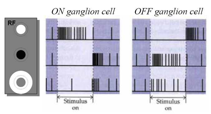

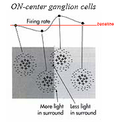
Figure 7: Firing Rate of On-center Ganglion Cells
It can be concluded that from figure 7, the ganglion cells itself can be modeled using a neural network with the input weights carefully designed to model the center-surround characteristic of the ganglion. From the graph in figure 7, it seems that the surround region carry negative weights and the center region carry positive weights, and when the center region is lighted, and partially surrounds lighted results in an increase in firing rate and when both are lighted, there is no increment in firing rate. [3]
Architecture Visual Pathway Outer Nuclear Layer Inner Nuclear Layer Ganglion Cell Layer