A.2 Gross Structure of the Brain
The human brain consists of two halves (hemispheres) and resembles a peeled walnut. These two hemispheres communicate with each other by a thick bundle of fibers called the corpus callosum. Although the two hemispheres seem to be mirror images of each other, they are different. The right hemisphere controls the left side of the body and vice-versa. Each hemisphere is covered by a thick layer of gray substance, the Cerebral Cortex. To increase the surface area, the cerebral cortex is heavily folded. The folds are called convolutions, or gyri, and the grooves are referred to as sulci, or fissures, if they are very deep. There are two main sulci (or fissures) in the bran, visible on the lateral surface. These are
the lateral sulcus (also know as the Sylvian fissure) and the central sulcus.
A.2.1 Cerebral Structures
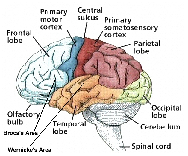
Each cerebral hemisphere can be divided into four lobes (frontal, parietal, temporal, and occipital) each of which specializes in different functions (Figure A.2).
Frontal Lobe:
The frontal lobes lie directly behind the forehead. The frontal lobe extends from the central sulcus to the anterior limit of brain. The central sulcus separates the frontal lobe and the parietal lobe. Inferiorly, the frontal lobe is separated from the temporal lobe by the sylvian fissure which is also called the lateral fissure. It contains the motor cortex and prefrontal cortex. The precentral gyrus, which may also be called the primary motor area or, most commonly, the motor strip is immediately anterior to the central sulcus. The amount of tissue on the precentral gyrus that is dedicated to the innervation of a particular part of the body is proportional to the amount of motor control needed by that area, not just its size. For example, much more of the motor strip is dedicated to the control of the articulators than to the legs. The premotor area or supplemental motor area is immediately anterior to the motor strip. It is responsible for the programming for motor movements. It does not, however program the motor commands for speech as these are generated in Broca's area which is also located in the frontal lobe. Broca's Area is found on the inferior frontal gyrus in the hemisphere that is dominant for language. The most anterior part of the frontal lobe is (called the prefrontal cortex) and is involved in complex cognitive processes like reasoning and judgment.
Parietal Lobe:
The Parietal Lobe is immediately posterior to the central sulcus. It is anterior to the occipital lobe, from which it is not separated by any natural boundary. Its inferior boundary is the posterior portion of the lateral fissure which divides it from the temporal lobe. The Parietal region processes body information including touch, information from muscle stretch receptors and joint receptors. The postcentral gyrus which is also called the primary sensory area or the sensory strip is immediately posterior to the central sulcus. This area receives sensory feedback from joints and tendons in the body and is organized in the same manner as the motor strip. The sensory association areas are located behind the postcentral gyrus. These areas are capable of more detailed discrimination and analysis than is the primary sensory area. They might, for example, be involved in sensing how hot or cold something is rather than simply identifying it as hot or cold. Information is first processed in the primary sensory area and is then sent to the secondary sensory areas.
Temporal Lobe:
The temporal lobe is located laterally in each hemisphere, near the temples. The Temporal Lobe is inferior to the lateral fissure and anterior to the occipital lobe. It is separated from the occipital lobe by an imaginary line rather than by any natural boundary. The temporal lobe is associated with auditory processing, olfaction, and some complex aspects of vision (i.e., perception of complex patterns and faces). It is also involved in semantics, or word meaning. Wernicke's Area is located on the posterior portion of the superior temporal gyrus. In the hemisphere that is dominant for language, this area plays a critical role in the ability to understand and produce meaningful speech. The anterior transverse temporal gyrus, is the primary auditory area. There are two secondary auditory or auditory association areas which make important contributions to the comprehension of speech. They are not completely responsible for this ability, however, as many areas, including Wernicke's area, are involved in this process. The angular gyrus lies near the superior edge of the temporal lobe, immediately posterior to the supramarginal gyrus. It is involved in the recognition of visual symbols. Fibers of many different types travel through the angular gyrus, including axons associated with hearing, vision, and meaning.
Occipital Lobe:
The occipital lobe is located in the posterior, caudal end of the cortex. It is the main target for axons from thalamic nuclei that receive inputs from the visual pathways. It contains the primary visual cortex. The secondary visual areas integrate visual information, giving meaning to what is seen by relating the current stimulus to past experiences and knowledge. A lot of memory is stored here. These areas are superior to the primary visual cortex. It is important to remember that while some functions can be localized to very specific parts of the brain, others cannot be classified in this way because many areas are involved in their performance. Word-finding, for example, is associated with several different areas. Also, we cannot say that all higher level cognitive functioning is associated with the frontal lobe; the processing of word meaning carried out by Wernicke's certainly involves a sophisticated type of cognition. Also, right hemisphere lesions often result in cognitive/perceptual problems.
A.2.2 Subcortical Structures
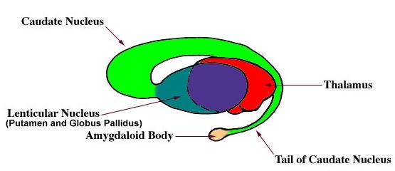
The Basal Ganglia are groups of neurons positioned subcortically. They include the caudate nucleus, putamen, and globus pallidus. The caudate nucleus and the putamen together form the corpus striatum or simply striatum. The globus pallidus contains more myelinated fibers than the adjacent striatum (putamen) and is accordingly, lighter in colour. The globus pallidus and the putamen together form a lens-shaped mass called the lenticular or lentiform nucleus. The caudate nucleus is an elongated C-shaped nuclear mass, which wraps around the upper and lateral border of the lateral ventricle. The putamen lies laterally to the globus pallidus. The amygdala, which is involved in emotion, was once classified as part of the basal ganglia, but is no longer categorized in this way. It is still considered to be a part of the limbic system. It is attached to the tail of the caudate nucleus. The subthalamic nuclei and the substantia nigra are both functionally related to the basal ganglia, but are not considered to be part of that structure. The corpus callosum, which is Latin for "large body" is the major group of commissural fibers. It is located some distance down inside the longitudinal cerebral fissure, the split that separates the hemispheres. The other two groups of commissural fibers are called the anterior commissure and the posterior commissure. Both are connected to the corpus callosum. The limbic system consists of both cortical and
Subcortical structures which are located on the medial, inferior surfaces of the cerebral hemispheres. The cortical areas classified as part of the limbic system include the hippocampus, the cingulate gyrus, and the subcallosal gyrus. The hippocampus, is a gyrus found on the medial edge of the temporal lobe. It is named for its shape, as hippocampus literally means "sea horse". The cingulate gyrus is immediately superior to the corpus callosum. The subcallosal gyrus is immediately inferior to the corpus callosum. The thalamus has been described as the switchboard for the cortex. It receives information from the cerebellum, the basal ganglia and from all sensory pathways with the exception of the
olfactory tract; it integrates the messages and sends them on to the cortex for further processing. Both the thalamus and hypothalamus are located in the center of the brain at the level of the temporal lobe. They are very well protected in this area. The thalamus is located below the caudate nucleus and the fornix and is medial to the lenticular nucleus. It is composed of two bodies which are separated from one another by the third ventricle, with one lying in each hemisphere. The two thalamic bodies are connected to one another by another part of the thalamus, the massa intermedia or thalamic adhesion, which makes up part of the ventricle. The subthalamus is located ventral to the thalamus and is important for motor movement. It has connections to the basal ganglia, thalamus and brainstem. The hypothalamus is a solid structure that is located immediately inferior to the thalamus. Part of it is also anterior to the thalamus. It forms the floor and part of the lateral walls of the third ventricle.
A.2.3 Cerebellum
Cerebellum is situated at the bottom of the brain. It consists of two cerebellar hemispheres joined in the midline by a narrow worm like portion called the vermis. Like the cerebral hemispheres, the cerebellum is covered with a layer of gray substance and is called the Cerebellar Cortex.