The functional cellular unit of the Central Nervous System is a nerve cell or Neuron. The neuron consists of a cell body (soma), dendrites attached to the body, and an axon. Many axons are surrounded by a myelin sheath, which has a white colour. Consequently those parts of the brain that consist of mainly myelinated axons are called white matter. The parts that contain aggregations of nerve cell bodies have a gray colour, hence the term gray matter. Groups of nerve cell bodies in other parts of the brain are called nuclei, or columns if they occur in long rows. Accumulations of nerve cell bodies outside the CNS (central nervous system) are called ganglia.
Directions and Planes:
There are a number of special words that are used to describe the position and direction of brain structures. These words help describe the location of structures relative to other structures. For example, we can say that the frontal lobe is �rostral� to the occipital lobe. Table A.1 gives a listing of the anatomical terminology used to describe directions.
|
Direction |
Description |
Direction |
Description |
| Ventral |
Toward the belly (front) |
Dorsal |
Toward the back |
|
Rostral |
Toward the nose |
Caudal |
Toward the tail |
|
Superior |
Toward the top (of the head/body) |
Inferior |
Toward the bottom |
|
Lateral |
Away from the middle |
Medial |
Toward the middle |
|
Ipsilateral |
On the same side |
Contralateral |
On the opposite side |
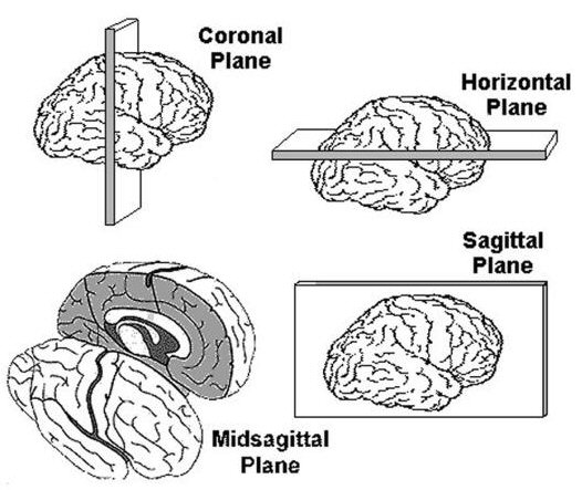
The brain, like all biological structures, is three dimensional. So, any point on or inside the brain can be localized on three "axes" or "planes" - the x, y and z axes or planes. The brain is often cut (sectioned) into pieces for further study. These slices are usually made in one of three planes: the coronal plane, the horizontal plane or the sagittal plane as shown in Figure A.1.
The 'Talairach' coordinate system specifies locations relative to their distance from the anterior commisure (AC). The AC is a small but easy to spot region, making it an ideal origin for the coordinate system. Each location is described by three numbers, each describing the distance in millimeters from the AC: X is the left/right dimension, Y is the posterior/anterior dimension, and Z is the ventral/dorsal dimension. Therefore, the position 0x0x0 is precisely at the AC, while -32x21x10 is left (32mm), anterior (21mm) and dorsal (10mm) from the AC. In this atlas the axial slices are referred to by their Z coordinate and coronal images are referred to by their Y coordinate. Again, it is important to stress that normalization strives to retain the unique features of each individual brain, and therefore Talairach coordinates are only approximate when comparing locations to other individuals.
The coronal plane, horizontal plane and sagittal plane are shown in the figure A.1. The coronal plane is also called the frontal plane. It cuts the brain into the anterior and posterior parts. The horizontal (transverse or axial) plane cuts the brain into top (dorsal) and bottom (ventral) parts. The sagittal plane divides the right and left side of the brain into parts. The midsagittal plane would divide the right and left sides of the brain into two equal parts.