FNA thyroid: medullary carcinoma


Drs. Prolla and Diehl's INTERESTING CASE OF THE MONTH October 2001 Case B

200x M-G-G staining, dispersed plasmacytoid cells pattern

500x M-G-G staining, cytoplasmic pink granules

500x M-G-G staining, amyloid globule

500x M-G-G staining, oval shaped cells with pink cytoplasmic granules

1000x M-G-G staining, pink cytoplasmic granules

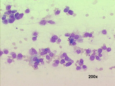
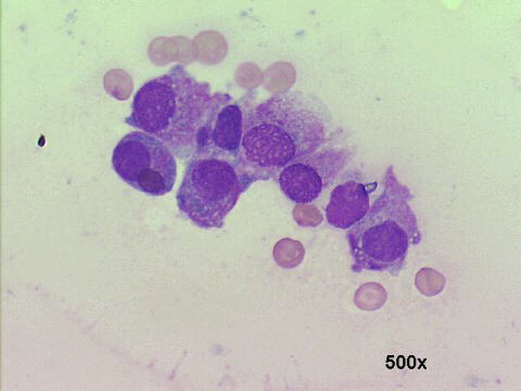
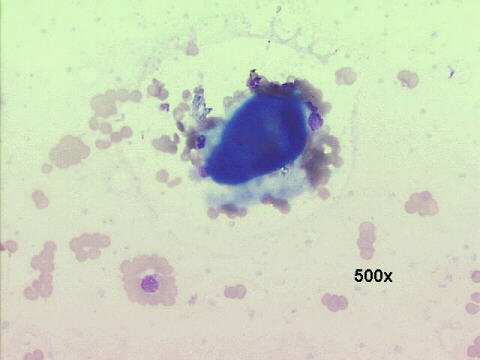
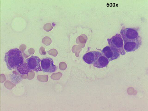
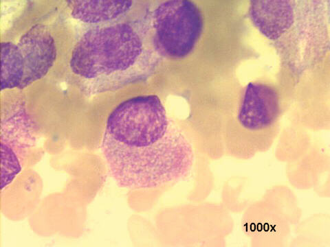
Cytology features: very cellular smears, with absent colloid, but some amorphous blueish material (probable amyloid), not to be confused with "chewing gum" colloid. The tumoral cells occur mostly as dispersed single cells, with ovoid or triangular shape. Their cytoplasm have a finely pink granular material. Histology revealed a typical medullary carcinoma of the thyroid. (H&E staining)Click here to see the histology pictures


Click here to see also Case A


References
List of cases
Hosted by www.Geocities.ws

1