|
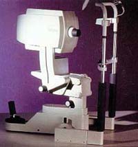
Heidelberg Retina Tomograph (HRT) |
|
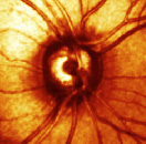
The established techniques for diagnosing and monitoring glaucoma include eye exams, photographs of the retina and vision field tests. Several problems exist in the current diagnostic methods. These include the need to dilate patients' pupils, difficulty of evaluating the retina through eye exams, and the fact that significant damage to the optic nerve may already have occurred by the time a vision field test detects glaucoma. A new type of diagnostic instrument called the Heidelberg Retina Tomograph (HRT) offers hope for improved detection and monitoring of glaucoma. HRT is a confocal scanning laser ophthalmoscope that generates three-dimensional images of the optic disc and peripapillary retina at the time of the patient visit. One can obtain topographic measurements of key parameters using HRT including retinal nerve fiber layer (RNFL) thickness, optic disc area and optic cup area (1). Previous studies have shown that HRT offers quantitative, objective and real-time analysis of the RNFL and does not require dilation of the pupils (2). Further studies have shown that HRT can discriminate between normal and glaucoma eyes with sensitivities and specificities of 78% and 91% respectively (3). Little information, however, is available on the ability of HRT to detect subtle changes in the retina over time such as those occurring in glaucoma patients. My goal is to use a promising analysis strategy for confocal scanning laser measurements, Probability Map Analysis, to determine if HRT can detect repeatable glaucomatous changes better than visual field exams. (1) Bathija, R, Zangwill, L, Berry, C, Sample, P, Weinreb, R. Detection of early glaucomatous structural damage with confocal scanning laser tomography. J of Glaucoma 7:121-127. 1998 (2) Dreher, AW, Tso, PC, Weinreb, RN. Reproducibility of topographic measurements of the normal and glaucomatous optic nerve head with the laser tomographic scanner. Am J Ophthalmology 111:221-229 1991 (3) Chauban, B, Blanchard, J, Hamilton, D, LeBlanc, R. Technique for detecting serial topographic changes in the optic disc and peripapillary retina using scanning laser tomography. Investigative Ophthalmology & Visual Science. 41:3:775-782. 2000
|