|
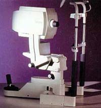
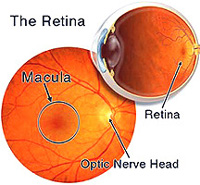
A patient's eye is scanned
with a confocal scanning laser called the Heidelberg Retina Tomograph (HRT)
and an image of the retina is produced. |
The image is three-dimensional and looks
like the picture above. The first picture taken is called the
baseline image. |
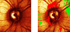
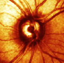
A year after the baseline image is taken,
our technicians obtain another image of the retina. The new image is
compared to the baseline image using analysis software which marks areas
of significant change as red or green.
Red = decrease in nerve layer thickness
Green = increase in nerve layer thickness
The two questions I want to answer are:
-
Do the red areas of change
correspond to glaucomatous nerve damage?
-
Can we use the laser
images to diagnose glaucoma earlier than the current diagnostic methods,
i.e. visual field exams ?
|