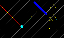
| Figure C1: Grey lines show the wavefronts of the incident X-ray beam. The red line shows the ray in the incident beam that scatters from the atom. The green line shows the scattered ray. The X-ray detector is shown in blue. The unimodular complex number C is plotted as a phasor arrow. Co is the value of C when the atom is at its equilibrium position. E = C - Co, is also plotted as a vector in the complex plane. 58c1.cpp 58c1.gif | |
|
Figure C2: In this case the motion of the atom does not
change the phase of the X-ray reaching the detector.
Therefore, this kind of motion does not cause inelastic
X-ray scattering towards the detector. 58c2.cpp 58c2.gif | 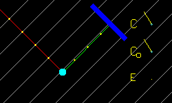 |
|
Figure C3: This is similar to Fig. C1 except that the
amplitude of the atom's motion is much less. This more
closely corresponds to the vibrations of a PMI where
the amplitude is much smaller than the X-ray or
neutron wavelength. 58c3.cpp 58c3.gif |  |
|
Figure C4: In this case two atoms both oscillate. Either
atom by itself would scatter X-rays inelastically towards
the detector, but together they almost cancel each other.
This is the essence of why torsional modes of PMI's
scatter X-rays so weakly. E(t) = Σj (Cj(t) - Coj ).
The small signal in E(t) is actually the 2nd harmonic, and is thus a
nonlinear effect. Actual PMI's have such slight motion that there
is no inelastic scattering of higher harmonics of the motion. 58c4.cpp 58c4.gif | 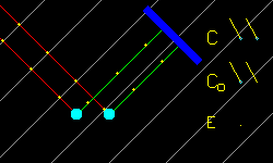 |
|
Figure C5: This illustrates how strong inelastic X-ray
scattering can take place from a spheroidal phonon
mode of a PMI. 58c5.cpp 58c5.gif | 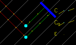 |