 |
| Fundus photographs of BRVO patients before and after vein decompression surgery |
|
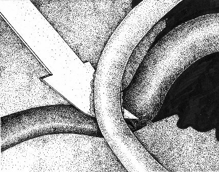
| Cutting the sheath between the artery and the vein. |
 |
 |
 |
 |
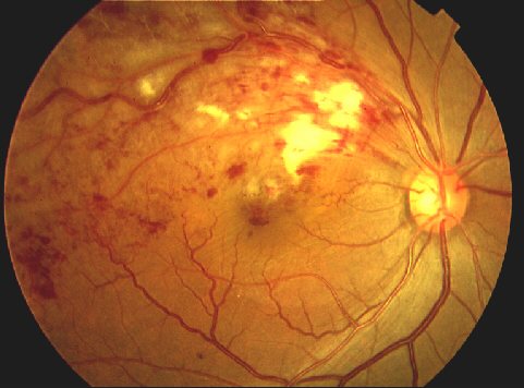
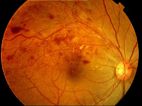
| Fundus photograph of patient before (left) and after (right) vein decompression surgery. Vision improved form 1/60 to 0.3 in 7 weeks. |
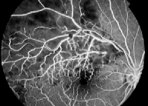
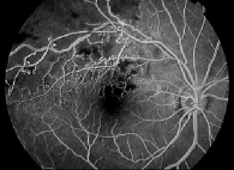
| Fundus fluorescein angiography of patient in figure 3 before (left) and after (right) vein decompression surgery showing an improvement in retinal blood circulation. |
| Fig. 1 |
| Fig. 3 |
| Fig. 4 |
| Fig. 5 |
 |
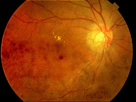 |
| Fundus photograph of patient before (left) and after (right) vein decompression surgery. Vision improved from 0.3 to 0.5 in 13 weeks after surgery. His surgery was made interesting by the fact that the occlusion was inside the cup of the optic disc. |
 |
| Fig. 2 |
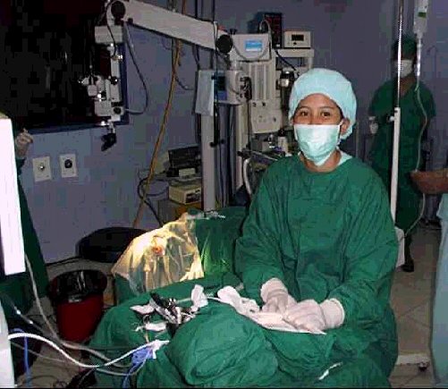
| Preparing for surgery |