Study of the potential for a combined
optical and x-ray mammography system using a compressed breast phantom
Breast Cancer and Mammography
- In UK, 40,000 new cases of breast cancer
are diagnosed each year
- Approximately 11,500 women die of breast
cancer each year in England and Wales
- The NHS Breast Screening Programme (NHSBSP)
invites women from the age of 50 to be screened for breast cancer
This screening involves a mammogram during which the patients breast is compressed
and an image is produced of it using low energy x-rays
The main drawback of mammography
is that it is unable to distinguish abnormalities as malignant or benign. This
means that any patient who is diagnosed with an abnormality will have to undergo
further testing.
Using diffuse optical tomography
(DOT) to detect breast cancer
- Near-infrared light is emitted into the
breast. Some of the light will be absorbed by areas of the breast such as
blood
- The transmitted light is detected and the
time of flight and intensity of the photons detected will be processed and images
produced from the data
- The images produced will identify tumours
as they are more vascular than normal breast tissue
The main drawback of using diffuse optical
tomography to detect breast cancer is that the images have poor spatial
resolution
Combining DOT with mammography
- X-ray images can be used to locate abnormalities
and provide information on their exact position and size
- Optical images can be used to classify abnormalities
as malignant or benign
- This would reduce the need for further testing
Aims of this project
- Design and construct compressed and uncompressed
breast phantom which can be used in both x-ray and optical imaging
- Image the compressed phantom using the two
techniques
- Combine the images
- Design a combined mammography system
Breast Phantoms
- Phantoms represent a part of the body such
as the breast by simulating its properties
- They are used in research and as quality
assurance tools
- Phantoms are typically made of epoxy resin,
acrylic and wax
Designing the phantoms
- The linear attenuation coefficient of the
optical phantom material (epoxy resin) used by the BORL
department of UCL was found experimentally
| |
µ (cm-1) at 60keV |
| Optical phantom material |
0.228 ± 0.006 |
| Normal breast tissue |
0.201 |
- This material was found to be suitable to
use as the normal breast tissue of the phantom
- To provide a suitable contrast between the
normal breast tissue and the tumours of the phantom, a difference comparable
to that of normal breast tissue and tumours had to be achieved
- Linear attenuation coefficient of epoxy resin
with talc and chalk found experimentally
- It was found that the addition of 5% talc
to the epoxy resin increased its linear attenuation coefficient by 0.011cm-1
- To achieve the required contrast, a difference
of approximately 0.015cm-1 at 60keV was needed
| |
Sample µ (cm-1)
at 60keV |
| Epoxy resin |
0.209±0.008 |
| Epoxy resin + talc (5%) |
0.220±0.008 |
- The optical properties of epoxy resin and
solutions with chalk and talc were found experimentally
- It was concluded that talc did not effect
the optical properties of the epoxy resin but chalk did
Final design
- Shape and thickness of the compressed breast
phantom based on a compressed breast
- The tumours in the phantom contain 10% talc
as this produces a sufficient contrast
- The absorption coefficient of tumours is
2.5 times that of normal breast tissue of the phantom
- 4 tumours of 2 different diameters were
set in the phantom at ¼ and ½ the depth of the phantom
- 2 markers were also put inside the phantoms
to be used to align images of the phantom. The markers contain 20% talc and
have optical properties 10 times greater than those of the normal breast tissue of the phantom
For details of the compressed breast phantom,
click here. The uncompressed breast phantom is
the same as the compressed except that it is double the thickness.
Images of the phantom
To see the x-ray image of the
phantom, click here
An example of the optical images obtained of
the phantom is shown here:
Double-sided
interface |
Single-sided
interface |
10mm |
20mm |
10mm |
20mm |
 |
 |
 |
 |
The double-sided interface images were obtained
when sources and detectors were above and below the phantom. The single-sided
interface images were obtained with all the sources and detector on one side
of the phantom. The images are shown at 10mm and 20mm below the top of the phantom
because these are the depths at which the middle of the tumours were set.
The images produced using the double-sided interface
were of better quality and provided more information. For this reason, the design
of the combined system is based on having sources and detectors above and below
the breast.
To see a selection of the combined images,
click here
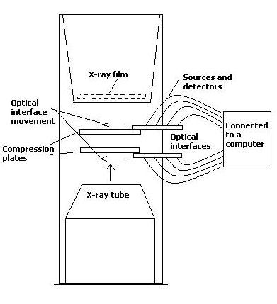
Design for a combined system
- The design is based on a standard mammogram
unit with the addition of a double –sided optical interface
- First an x-ray image is produced of the breast,
then, without releasing compression, the optical interfaces are moved onto the compression
plates so that the optical data can be acquired
- The compression plates would have to be
made of glass to reduce the interference in the optical images
Summary
- A compressed and an uncompressed
breast phantom have been produced
- The compressed phantom has
been imaged using x-ray mammography and DOT
- Sources/detectors above and
below phantom produced best optical images
- Combination possible
- Need improvements in the optical
image quality and a reduction in the optical data acquisition time before
the technique could be considered in routine diagnosis of breast cancer
Home
Abstract