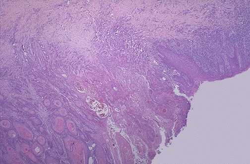
Cervix: Squamous cell carcinoma
Click to see other slides: [1]
A
50 yr old woman noticed post coital bleeding 6 months ago.
She now has intermittent spotting and vaginal discharge.
An ulcerated exophytic growth was seen in the cervix.
A PAP smear was taken followed bydefinitive surgery.
1.
What are the key histological features that indicate
Malignancy:
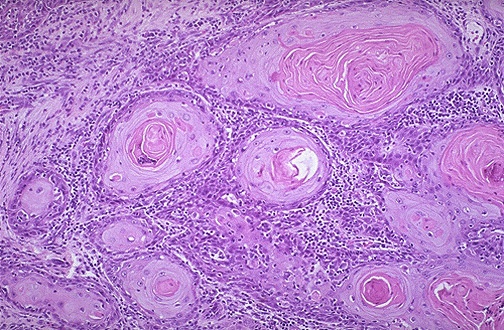
Sheets,
nests of atypical cells invading into the subepithelial stroma, basement
membrane is breached
Squamous
differentiation:
Keratin
pearls, sheets of atypical cells with pink cytoplasm
2.
How
does this lesion differ from CIN III?
Invasion
of subepithelial stroma
Basement
membrane breached
3.
What
do you expect to see in the cervical cytology smear?
Atypical
cells with high nucleocytoplasmic ratio, similar to that seen in CIN III.
4.
Account
for the presenting complaints.
Ulceration
and necrosis of exophytic tumour
Carcinoma
erodes into patent blood vessels, accounting for post coital bleeding
<< PREVIOUS INDEX NEXT SLIDE >>
Copyright � Joseph Ong 2003