Cervix: CIN III
Click to see other slides: [1]
A
35 yr old woman who had history of multiple sex partners in the past was found
on routine gynaecological check up to have an abnormal PAP smear.
She was other wise asymptomatic.
Biopsy of the cervix was performed.
1.
What is CIN and how is it classified?
CIN
�
Cervical intraepithelial neoplasia
Classified
into CIN I, II, III based on the level of dysplasia of the cervical epithelium
CIN
I �
mild dysplasia
CIN
II �
moderate dysplasia
CIN
III �
severe dysplasia / carcinoma in situ
2.
Describe
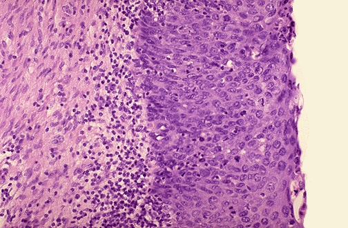
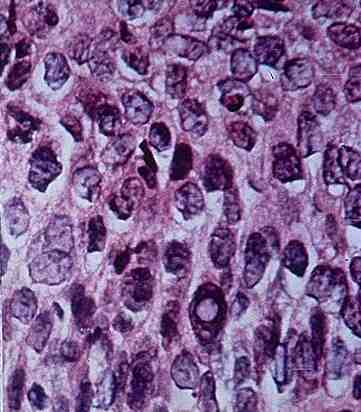
the key histological features of the biopsy which shows CIN III.
Atypical
cells in entire thickness of cervical epithelium
Increased nucleocytoplasmic ratio
Hyperchromatic nuclei
Variation in nuclear size
Increased abnormal mitoses
Loss of polarity
Loss
of surface maturation / differentiation
Basement
membrane intact
No
invasion of epithelial cells into the stroma
3.
How
do you distinguish CIN III from invasive squamous cell carcinoma of the cervix
histologically?
CIN
�
basement membrane intact and no invasion into the stroma
SCC
�
basement membrane breached,
malignant epithelial cells found in stroma
4.
Try
describing the cytological findings in the abnormal PAP smear by extrapolating
from your observation of the cervical biopsy.
Pleomorphic
cells with high nucleocytoplasmic ratio
Total
lack of surface differentiation as the exfoliated cells originated from surface
epithelium
5.
What
does �PAP� stand for?
Is PAP smear equivalent to cervical smear?
PAP � Papanicolaou.
Yes.
6.
What
is the significance of the social history?
The
history of first intercourse of an early age, multiple sex partners, high risk
male sex partners are all associated with increased risk for cervical cancer and
precancerous dysplasia
7.
How
do cases of CIN usually present?
Generally
asymptomatic, shedding of abnormal cells from the cervix is usually detected by
routine PAP smear
<< PREVIOUS INDEX NEXT SLIDE >>
Copyright � Joseph Ong 2003