Liver: Hepatocellular carcinoma
Click to see other slides: [1]
A
70-yr-old man who had been losing much weight lately was found to have an 8 cm
diameter space-occupying lesion in the right lobe of his liver.
1.
Name some causes of space-occupying lesions in the liver.
Cavernous
hemangiomas
Actinomycosis
Liver
abscess
Primary
carcinoma (hepatocellular, cholangiocarcinoma)
Metastatic
carcinoma
2.
What is your diagnosis in this case?
Primary
hepatocellular carcinoma
3.
What
associated liver conditions are likely to be present?
Cirrhosis.
4.
What
pre-operative blood tests would have been done for the following purposes and
why?
a)
Diagnosis
Alpha
fetoprotein level (commonly in xs of 400ng/ml in patients with hepatocellular
carcinoma)
b)
Assessment of fitness for major surgery
ALT,
AST, GGT levels
Coagulation
time e.g. PT, PTT
5.
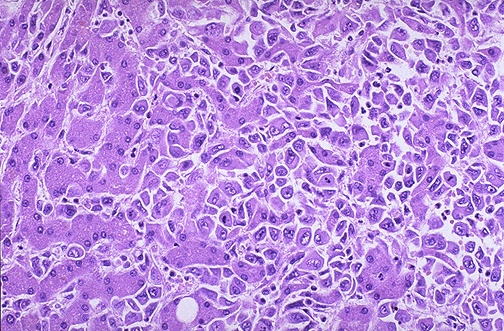
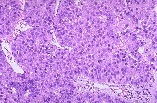
List
the diagnostic histological features.
Trabeculated
(thickness > 1 cell thick)
Presence
of sinusoids (lined by normal endothelial cells)
Eosinophilic
cytoplasm, hyperchromatic nuclei with prominent nucleoli
Large
number of mitoses
Increased
nuclear to cytoplasmic ratio
Bile
production
6.
What
pathological (gross and microscopic) parameters have prognostic implications?
Staging
- T (size of primary tumour)
- N (nodal involvement)
- M (metastases)
Cirrhosis
Number
of tumours
7. True / False MCQs:
The
following are characteristic of this condition:
Bile production
T
Alpha-fetoprotein production
T
Tumour embolisation to the lung T
Intraperitoneal haemorrhage T
Mucin production
F
<< PREVIOUS INDEX NEXT SLIDE >>
Copyright � Joseph Ong 2003