MEDICAL IMAGING OF CEREBROVASCULAR DISEASE
by Ken McCormick, M.Ed., R.T.(R)(CV)
|
MEDICAL IMAGING OF CEREBROVASCULAR DISEASE by Ken McCormick, M.Ed., R.T.(R)(CV) |
|
|
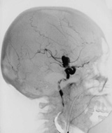
Welcome to the Cerebrovascular Disease program home page. Cerebrovascular disease (CVD), also known as stroke, is the 3rd leading cause of death in the U.S. At the end of this program of study you will be able to identify current mortality and morbidity statistics, distinguish between the different types of CVD, identify characteristics and causes for the different types of CVD, discuss patient management and appropriate treatments for acute stroke patients, identify cerebrovascular anatomy, and discuss the roles of various medical imaging modalities in the diagnosis and treatment of CVD. This page contains an outline of the topic areas contained in this program of study, with highlighted links to each unit. You may use this page as a central "jumping off" point to access any page in the program. The program consists of three main topic areas, a glossary, appendix, and self-test. The topic areas are arranged so you may begin with Unit 1 and proceed sequentially through the subsequent units. At the first of each unit, links are provided so that you may go immediately to a desired section. At the bottom of each page is an arrow box that allows you to go back to the previous page or forward to the next unit. You may access the home page, glossary or test from any page in the program. When you have completed the program and submitted your test answers, please fill out the online survey. Your input will help me to improve and enhance the learning experience offered by this program. Thank you! |
|
CONTENT OUTLINE |
|
|
|
|
|
|
|
Test |
|
Online Survey |
|
Return to Special Procedures Menu Page |
|
ACKNOWLEDGEMENTS Many of the images in this program were acquired through the courtesy of local technologists and physicians. I would like to thank the following individuals who contributed their efforts to this program: Alan Nooner, R.T.(R)(CT) and Annette Walters, R.V.T., of St. Joseph's Regional Health Center, Billy Shuffield, R.T.(R)(CV) of National Park Medical Center, Mark Parker, R.T.(R)(MR) of Hot Springs MRI Center, and for the excellent images from their respective modalities, and to Pete Barger, R.T.(R)(CT) of St. Joseph's Regional Health Center for his valuable assistance in scanning the images. I would also like to thank Drs. Cecil Cupp, Michael Hickman, Mark Robbins and Phillip Smith of Hot Springs Radiology Services, Ltd., for their generous assistance with image interpretation. |
|
NEXTÞ |
Ó
Images and text copyright Ken McCormick, April 1999. All rights reserved.