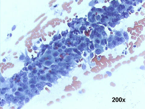
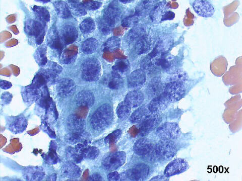
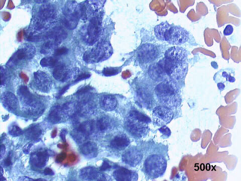
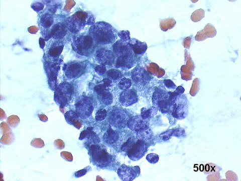
The smear shows abnormal glandular cells in a palisading nuclear arrangement with crowded overlapping nuclei protruding from the periphery of the cell clusters (feathering). Nuclear enlargement and stratification are evident. Hyperchromasia with moderately granular chromatin is also evident. Clean background without inflammatory cells or debris. These findings strongly suggest adenocarcinoma in situ of endocervix, a diagnostic category reinstated in The 2001 Bethesda System. A cone biopsy proved the cytological diagnosis.
The July 2002 Case of the month of the Australian Society of Cytology is a case of cervical endometriosis. See it for this difficult differential diagnosis.