Drs. Prolla and Diehl's INTERESTING CASE OF THE MONTH November 2006 case B
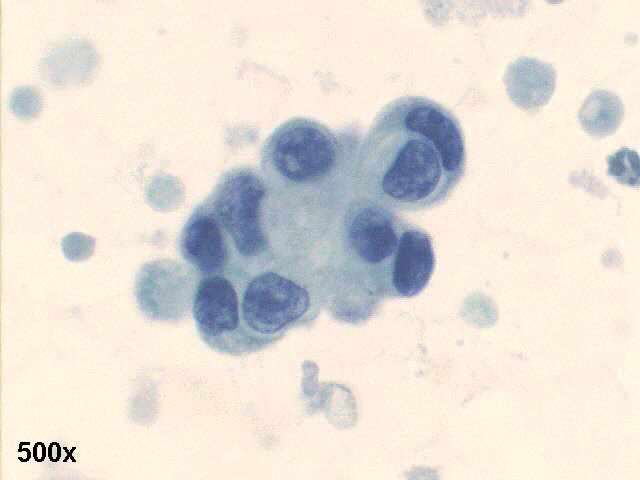
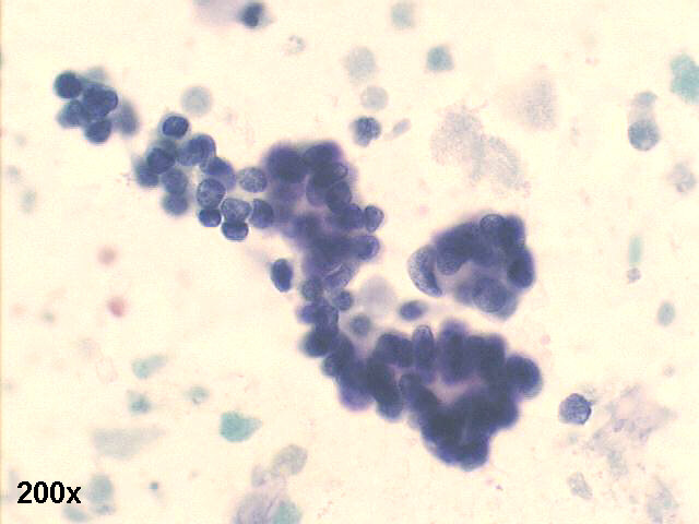
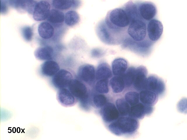
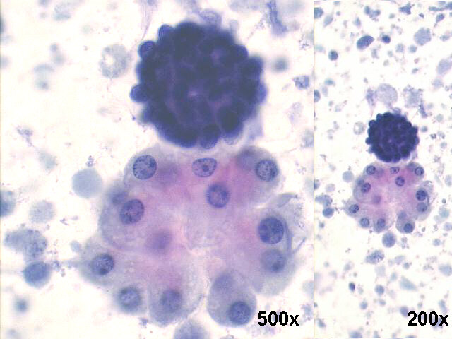
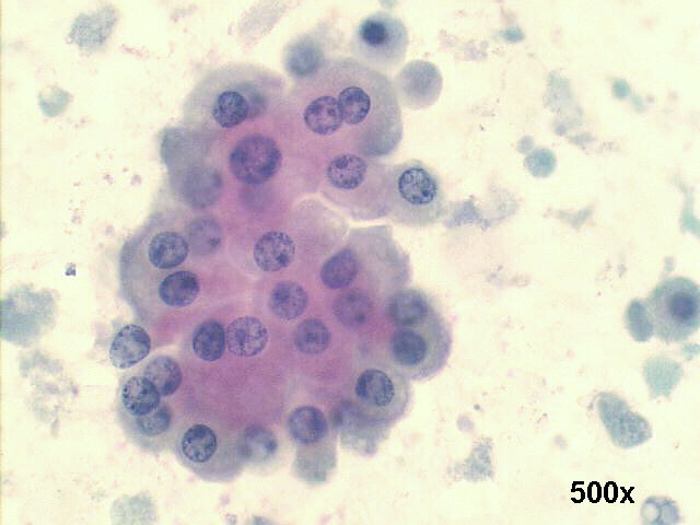
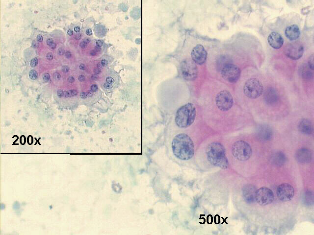
Left breast bloody nipple discharge 50-year old female: intraductal papilloma with apocrine metaplasia

500x, Pap. staining, cluster of atypical cells

200x, Pap staining, pseudo-papillary fragment

500x Pap staining, pseudo-papillary fragment, detail view shows atypical cells with high N/C ratio and hyperchromasia

200x and 500x Pap., a hyperchromatic epithelial cell ball, and a cluster of benign looking metaplastic apocrine cells.

500x Pap staining, a cluster of benign looking metaplastic apocrine cells, and  scattered macrophages in a proteinaceous background.

200x and 500x Pap staining, and a cluster of benign looking metaplastic apocrine cells in a proteinaceous background.

The smears show a double population of cells: groups of benign looking metaplastic apocrine cells, and pseudo-papillary groups of atypical hyerchromatic cells, in a background of proteinaceous debris with scattered macrophages. This pattern suggests an intraductal proliferative lesion, possibly an intraductal papilloma (rule out intraductal papillary carcinoma), and an associated apocrine metaplasia.
Spontaneous bloody nipple discharge is worrisome for two conditions: intraductal papilloma (about 85~90% of the cases of bloody nipple discharge) and intraductal or other types of breast cancer (about 10~15% of bloody nipple discharge).
The surgical follow up revealed an intraductal papilloma just below the left breast nipple area, with ductal ectasia, and some areas of apocrine metaplasia.

Case A November 2006 Case B November 2006 References List of cases Atlas
Hosted by www.Geocities.ws

1