Drs. Prolla and Diehl's INTERESTING CASE OF THE MONTH November 2008 case A
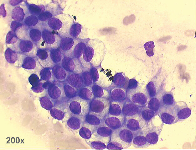
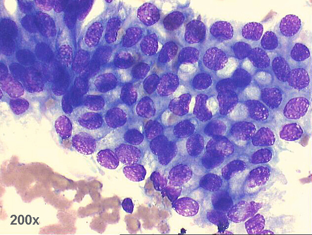
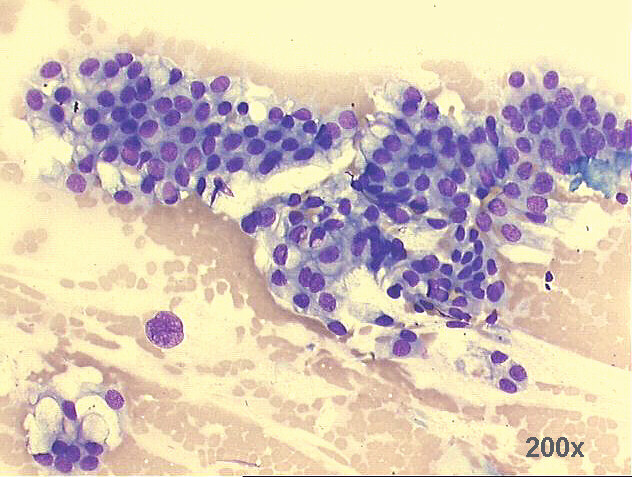
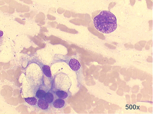
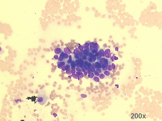
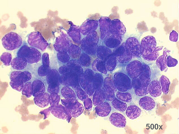
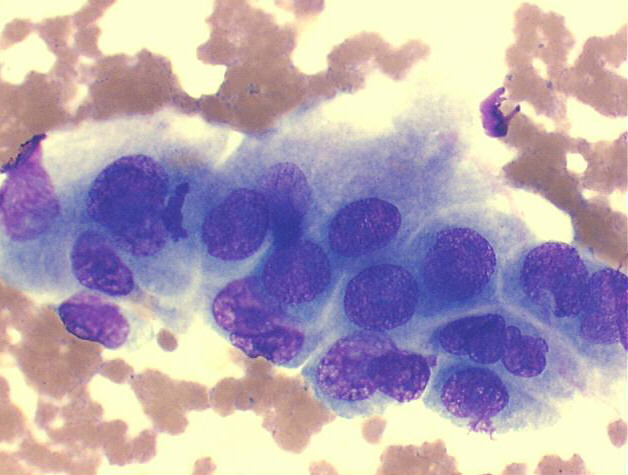
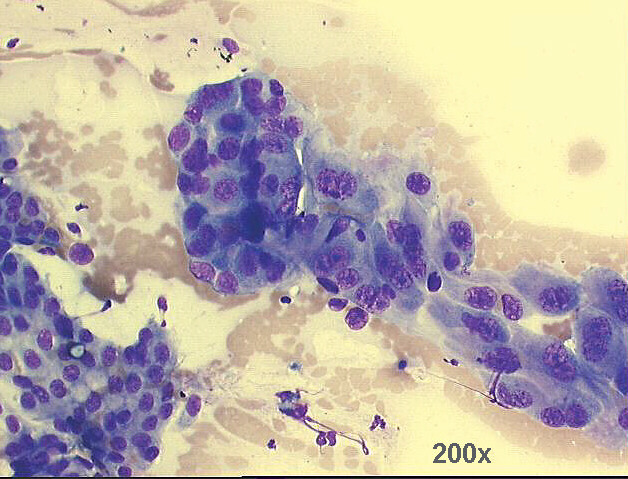
Endoscopic gastro-esophageal brushings, 78-year old female with past history of Barrettīs esophagus: adenocarcinoma

500x M-G-G staining - group of columnar and goblet cells, with benign morphology.

500x M-G-G staining - another group of columnar and goblet cells, with benign morphology.

200x M-G-G staining - low power view of groups of columnar and goblet cells, with benign morphology. Notice a very large naked nucleus.

500x M-G-G staining, high power view of previous image: detail of the large naked nucleus, very suspicious of malignancy

200x M-G-G staining - group of glandular cells, with morphology suggestive of malignancy.

500x M-G-G staining, high power view of previous image: malignant looking cells, with crowding, hyperchromasia and anisonucleosis

500x M-G-G staining - another high power view: malignant looking cells, with crowding, hyperchromasia and anisonucleosis.

200x M-G-G staining - another group of glandular cells, with morphology suggestive of malignancy.<">

500x M-G-G staining, high power view of previous image; the nuclear abnormalities are obvious, with chromatin clumping and large irregular nucleoli

500x M-G-G staining, another high power view of previous image, also showing obvious nuclear abnormalities, with chromatin clumping and large irregular nucleoli


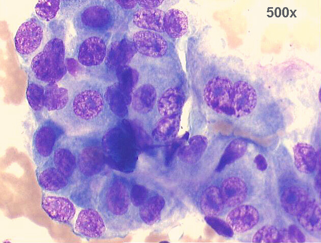
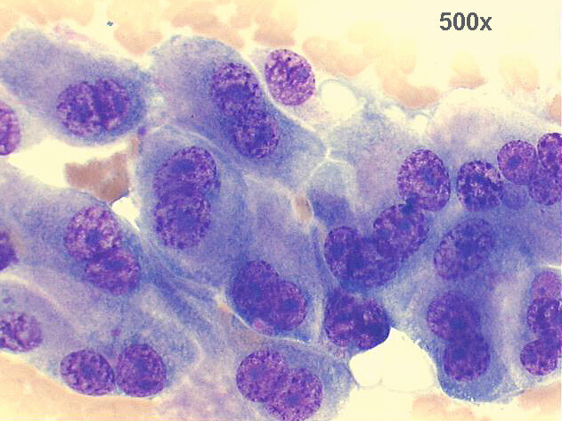
The patient had a history of Barrett's esophagus for several years. The smears show abundant number of columnar cells, with normal looking morphology and a second population of abnormal cells: loss of mucin production, and irregular groups of large cells with highly atypical nuclei suggestive of adenocarcinoma. This was confirmed by endoscopic biopsy, and also after surgical biopsies.


Case A November 2008 References List of cases Atlas
Hosted by www.Geocities.ws

1