Pleural fluid, 66-year old male, with large mediastinal mass:
Small cell carcinoma
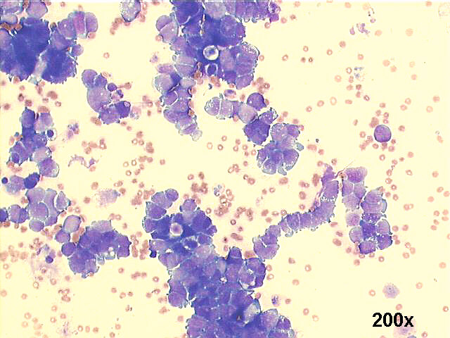
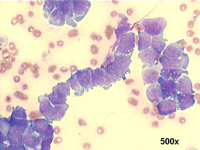
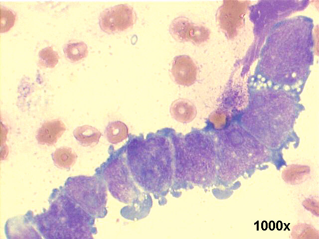
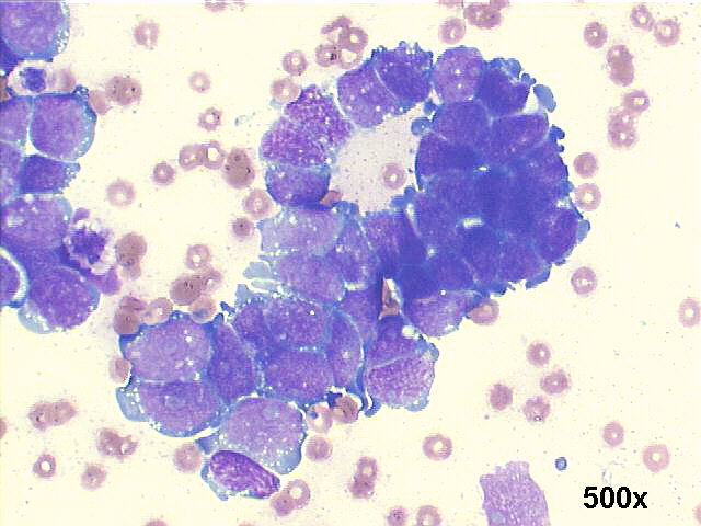
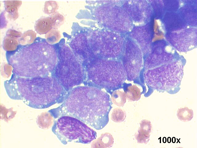
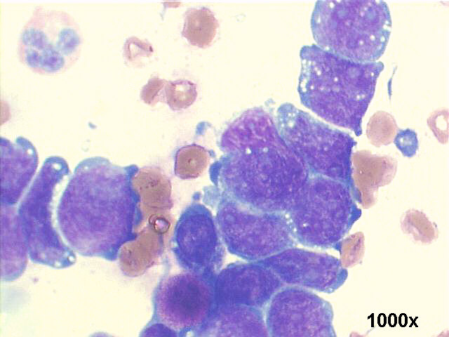
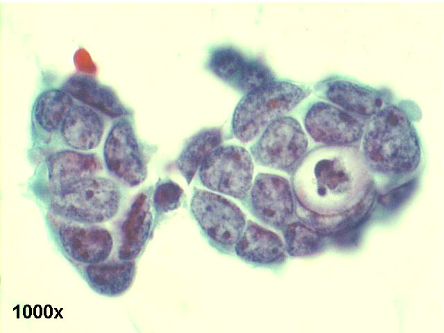
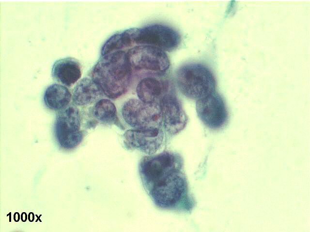

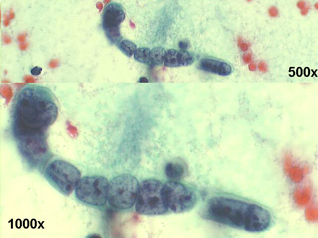
The smears show the typical features of small cell carcinoma: small cells, with some loose groups showing nuclear molding, many of them with tandem or indian file cell pattern. The cytoplasm is scant or absent. The nuclei are oval or angulated, with "salt and pepper" chromatin, small nucleoli, in the Papanicolaou staining. The M-G-G stained smears show many cellular details: nuclear molding, somewhat large blue nucleoli, very high N/C ratio, cytoplasmic projections, small clear vacuoles (both cytoplasmic and nuclear). Some people would call this an "intermediate type" of small cell carcinoma, to distinguish it from classical "oat cell" pattern, but the treatment and prognosis is the same. The DNA streaking or nuclear smudging ("crush artifacts") are absent in this case, as it is usual to be absent in serous fluids material.
Main features of Small cell carcinoma Cellular patterns: |
