Drs. Prolla and Diehl's Case of the month ~ October 2007

FNA 3.5cm in diameter left ovarian mass, 11-year old female: mature teratoma (ovarian dermoid cyst).

ovarian dermoid cyst 500x M-G-G staining, many skin squamous anucleated cells

ovarian dermoid cyst 200x M-G-G staining, many skin squamous nucleated and anucleated cells

ovarian dermoid cyst 500x M-G-G staining, many skin squamous nucleated and anucleated cells

ovarian dermoid cyst 200x M-G-G staining, many skin squamous nucleated cells

200x M-G-G staining, a sheet of benign mesothelial cells

ovarian dermoid cyst200x H&E staining, cross secion of many hairs on the wall of the cyst, and a foreign body giant cell.

ovarian dermoid cyst 500x H&E staining, detail view showing the cross section of hairs, and of the foreign body giant cell.

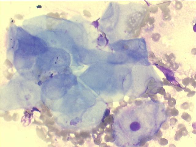
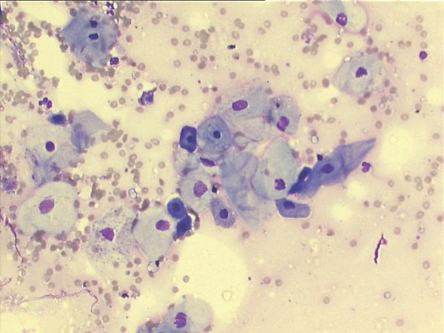
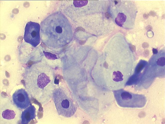
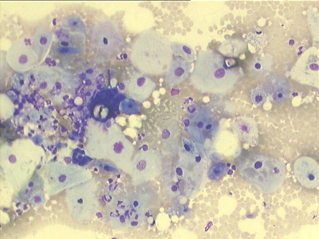
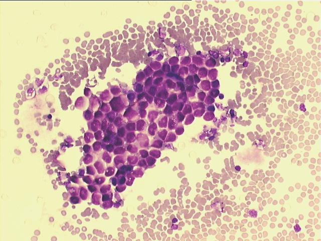
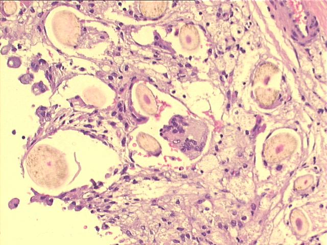
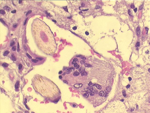
The radiologist informed that a hair came out from the needle while preparing the smears... This and the benign squamous cells, both anucleated and nucleated, point to the confirmed diagnosis of mature teratoma, or ovarian dermoid cyst. The other ovary showed also a benign teratoma, but with some less mature areas.


Case October 2007 References List of cases Atlas
Hosted by www.Geocities.ws

1