Drs. Prolla and Diehl's Case of the month ~ September 2007

FNA thoracic nodule, 60-year old female, BAC and papillary adenocarcinoma

BAC and papillary adenocarcinoma, many intranuclear cytoplasmic pseudovacuoles 500x M-G-G staining

BAC and papillary adenocarcinoma, many intranuclear cytoplasmic pseudovacuoles 500x M-G-G staining

BAC and papillary adenocarcinoma, 500x M-G-G staining

BAC and papillary adenocarcinoma, 500x Pap staining

BAC and papillary adenocarcinoma 500x Pap staining

BAC and papillary adenocarcinoma, many intranuclear cytoplasmic pseudovacuoles 500x Pap staining

BAC and papillary adenocarcinoma, many intranuclear cytoplasmic pseudovacuoles 500x Pap staining

BAC and papillary adenocarcinoma, many intranuclear cytoplasmic pseudovacuoles 500x Pap staining

BAC and papillary adenocarcinoma, CT thorax partially solid nodule 8mm diameter

BAC and papillary adenocarcinoma CT thorax

CT thorax, volume rendering 562 mm3

BAC and papillary adenocarcinoma CT thorax, Fine needle insertion

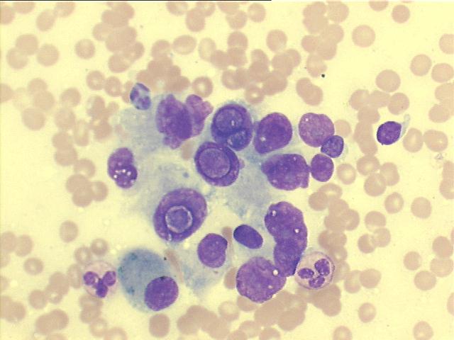
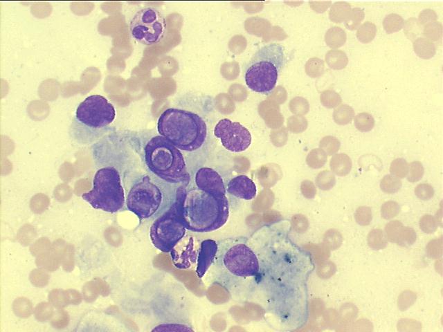
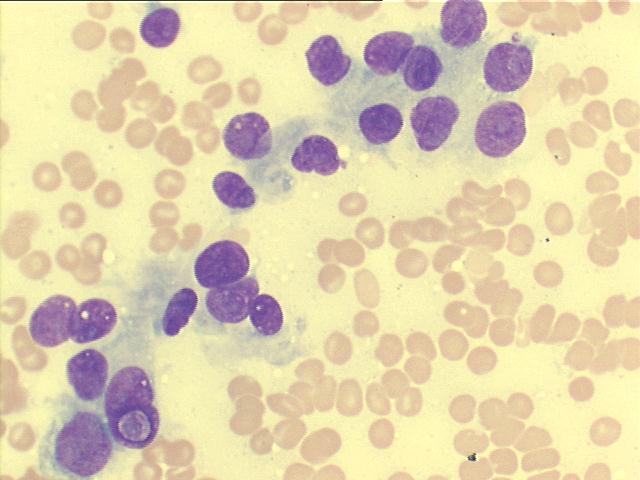
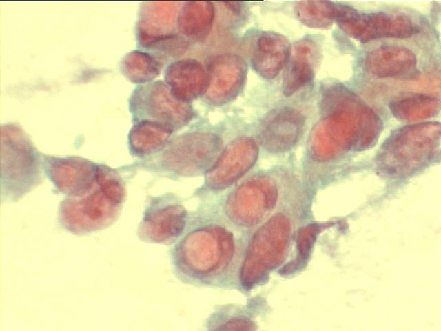
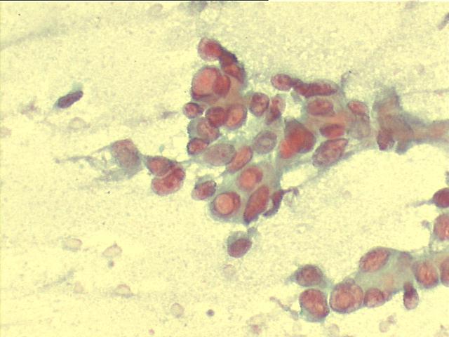
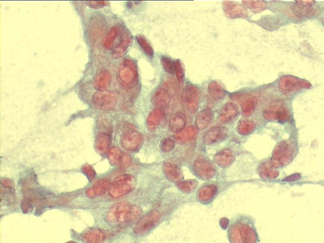
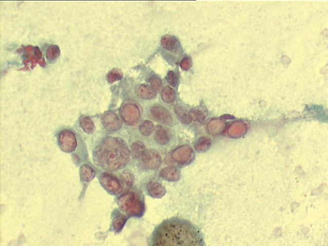
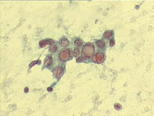
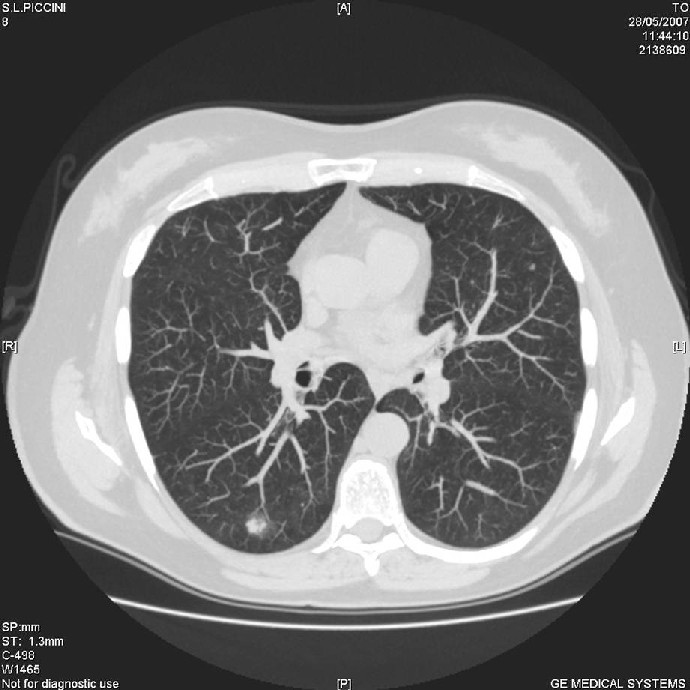
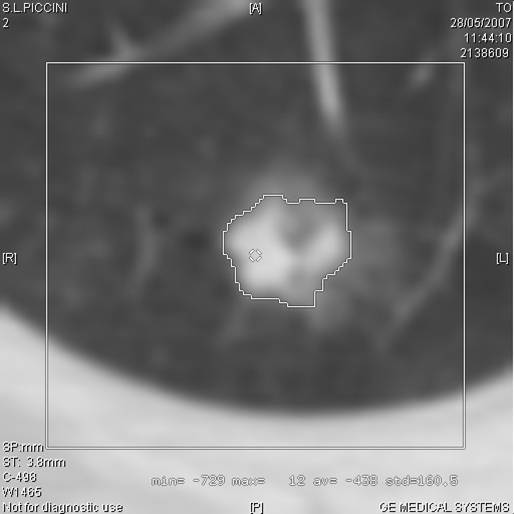
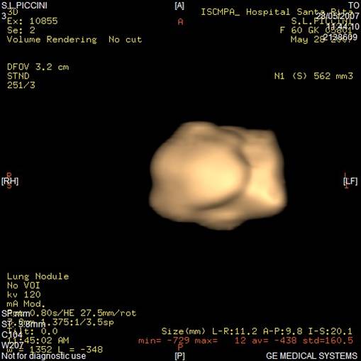
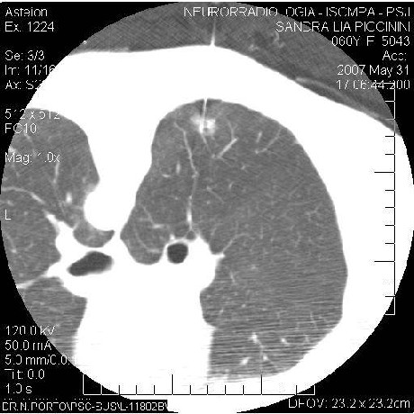
The cytology images show malignant cells with many intranuclear cytoplasmic pseudovacuoles, suggesting an adenocarcinoma of papillary type, and the CT images suggest a mixed BAC pattern, with the ground glass opacification with a solid component. The surgical specimen showed a papillary adenocarcinoma with a bronchoalveolar carcinoma (BAC) component.


Case A September 2007 References List of cases Atlas
Hosted by www.Geocities.ws

1