Drs. Prolla and Diehl's INTERESTING CASE OF THE MONTH July 2009
Cervico-vaginal cytology, 55-year old female.
Local recurrence well differentiated squamous cell carcinoma, previously treated with radiation therapy

Medium power view shows numerous squamous cells, many of them abnormal, 200x Pap staining

High power view: dysplastic squamous cells with anisocytosis and anisonucleosis including keratinization are diagnostic of invasive squamous cell carcinoma.
500x Pap staining

Dysplastic squamous cells with anisocytosis and anisonucleosis including keratinization are diagnostic of invasive squamous cell carcinoma. 500x Pap staining

Centrally located abnormal nuclei and flat arrangement of keratinizated cells is consistent with squamous cell carcinoma.500x Pap staining

Centrally located abnormal nuclei and flat arrangement of keratinizated cells is consistent with squamous cell carcinoma. 500x Pap staining

Dysplastic squamous cells with anisocytosis and anisonucleosis including keratinization are diagnostic of invasive squamous cell carcinoma. 500x Pap staining

Medium power view: less obviously dysplastic cells. 200x Pap staining

High power view: the malignant nature of the cells is not obvious. 500x Pap staining

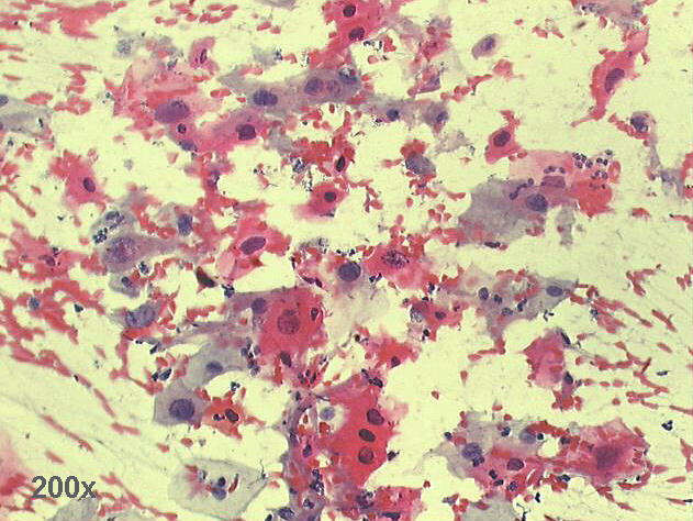
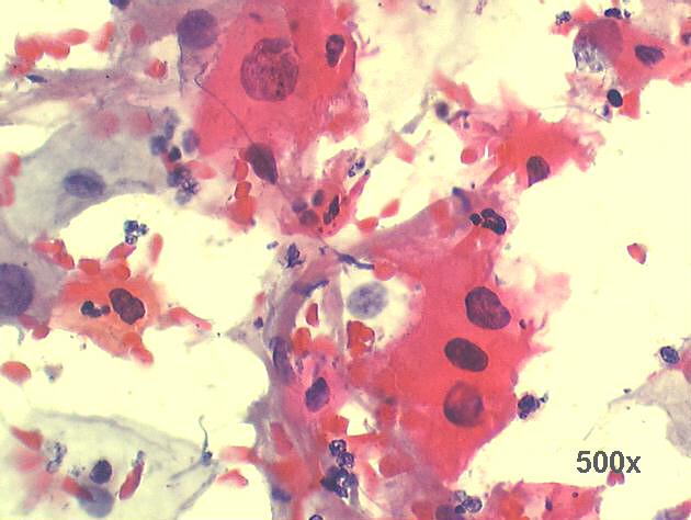
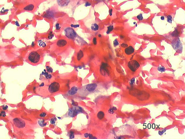
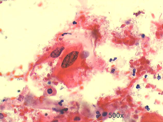
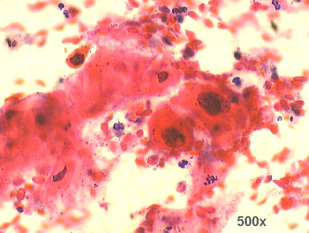
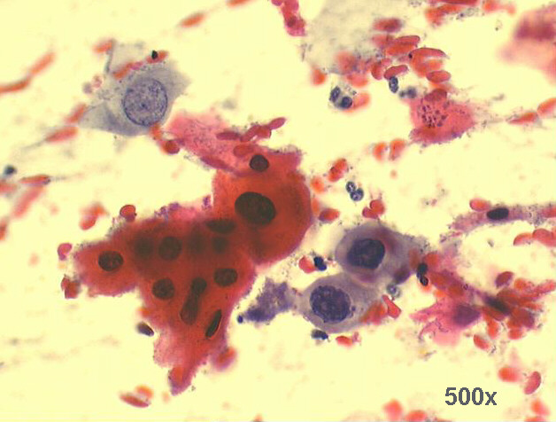
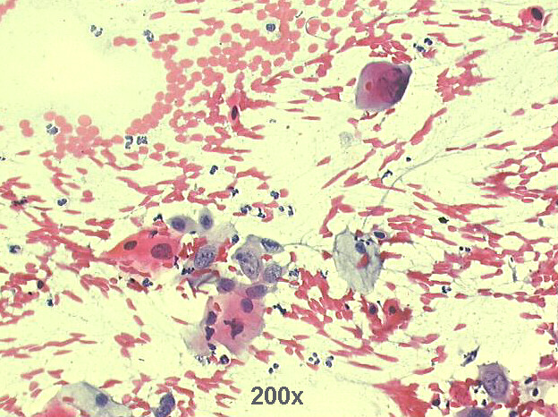
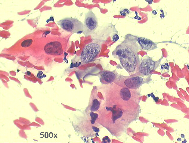
The smear shows abundant cellularity, with anisocytosis and anisokaryosis, many flat keratinized cells, with centrally located highly hyperchromatic nuclei: the cytological diagnosis should be local recurrence of well differentiated squamous cell carcinoma of the cervix, previously treated with radiation. This diagnosis was confirmed by the biopsy taken at the vaginal cupula.


Back to Case July 2009
List of cases
References
Atlas
Hosted by www.Geocities.ws

1