Drs. Prolla and Diehl's INTERESTING CASE OF THE MONTH June 2009
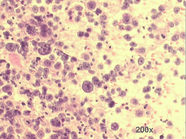
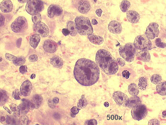
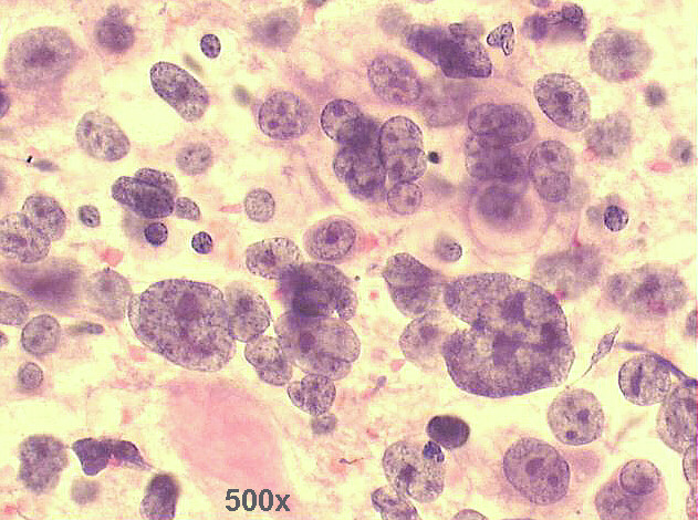
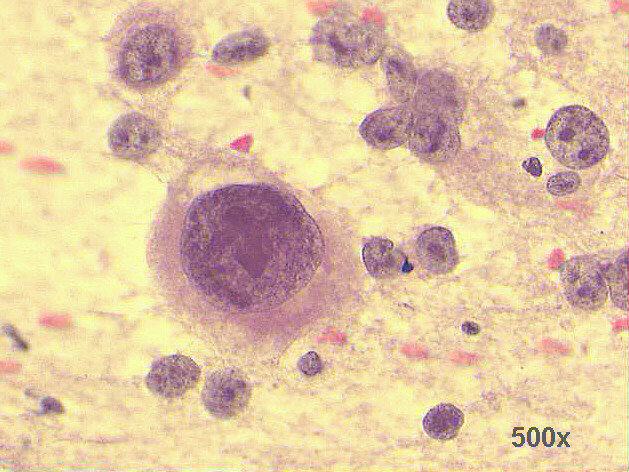
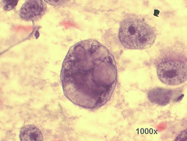
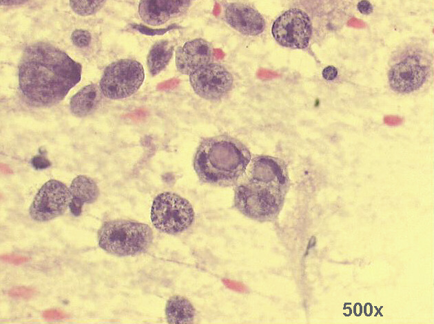
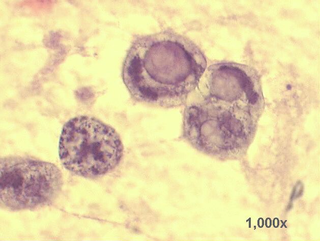
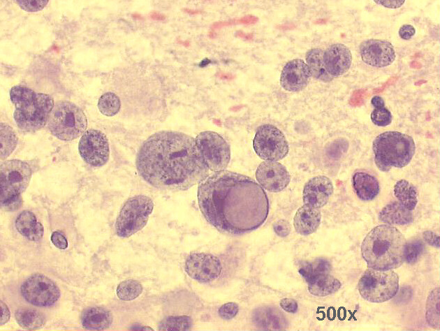
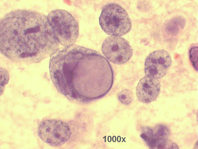
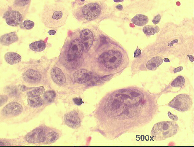
Left cervical node, transoperative imprint cytology, quick Pap staining, 67-year old male.
Metastasis from poorly differentiated squamous cell carcinoma

Medium power view: abundant cellularity of poorly differentiated malignant neoplasia, with preponderance of isolated cells and naked nuclei 200x Pap staining

High power view: quite anaplastic malignant isolated cells with large nucleoli and coarse chromatin 500x Pap staining

Another high power view: quite anaplastic malignant isolated cells with large nucleoli and coarse chromatin, predominance of naked nuclei 500x Pap staining

Quite large anaplastic cell with huge nucleolus 500x Pap staining

Huge naked nucleus with intranuclear vacuoles 1,000x Pap staining

Several anaplastic cells, some of them with intranuclear vacuoles 500x Pap staining

Detail view of the two cells with intranuclear vacuoles 1,000x Pap staining

Anaplastic cell with large intranuclear vacuole giving the cell a signet ring appearance 500x Pap staining

Detail view of the anaplastic cell with large intranuclear vacuole giving the cell a signet ring appearance  1,000x Pap staining

Only after careful scrutiny we found some groups of cells with epithelial appearance, like this one  500x Pap staining


I did a Pub Med search for imprint cytology used intra-operatively: I found 971 articles, with 58 reviews, as of July 21, 2009.
Most uses: sentinel node analysis in breast, head&neck tumors, veterinary medicine, lymphoproliferative lesions, quality control of frozen sections, central nervous system neoplasms. Most studies agree that it is a quick, highly accurate method, that can have clinical importance in helping the H&E studies, directing the immunehistochemical studies, and saving time and money in the definitive diagnosis of rare or unusual cases.
In this particular case, the technique helped to make intra-operatively a firm diagnosis of epithelial malignancy, rule out amelanotic melanoma. The H&E sections showed some areas of very poorly differentiated squamous cell carcinoma, and immunehistochemical studies confirmed the epithelial nature of the lesion, and ruled out melanoma and large cell lymphomas.


Back to Case June 2009
List of cases
Atlas
Hosted by www.Geocities.ws

Hosted by www.Geocities.ws

1