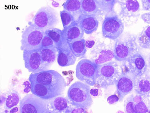
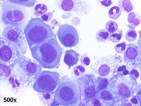
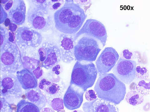
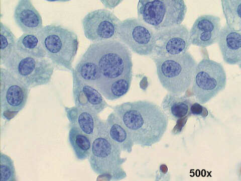
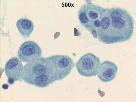
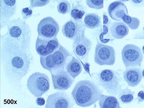
In his excellent book Malignant Effusions. A multimodal Approach to Cytologic Diagnosis(Igaku-Shoin, Tokyo, 1996) - chapter 4 - Serosal reaction to Injury, p.26-51, Dr. Carlos W M Bedrossian discusses at length the great variability of mesothelial cells. This is important to know, to help to distinguish malignant mesothelial cells from their benign reactive or hyperplastic variants, or from metastatic adenocarcinoma cells. In this case we see several morphological characteristics of those benign changes: doublets, triplets, or mosaics with windows between the cells ( caused by microvilli), multinucleation, anisonucleosis, cytoplasm with sharp ecto-endoplasmic demarcation, clasp-like connections, etc.
