Drs. Prolla and Diehl's
INTERESTING CASE OF THE MONTH April 2005
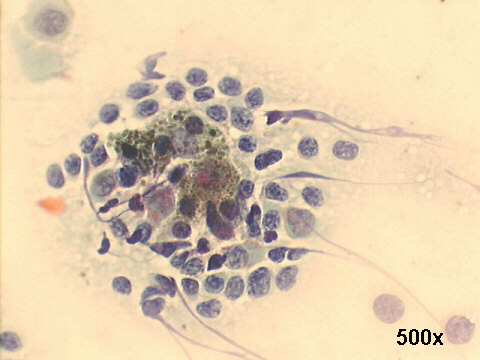
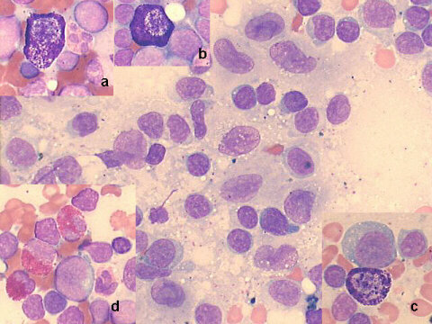
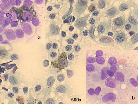
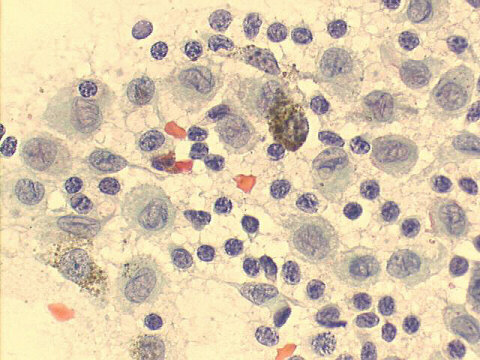
FNA cervical lymph node, 70-year old male: E.S. Hurwitt's dermatopathic lymphadenitis.
The smears show two main features: many histiocytes containing brown pigment (melanin, most probably), and the presence of many dendritic cells with longitudinal grooves, most likely Langherans cells. There are no malignant cells, making the diagnosis of metastatic melanoma unlikely. The Giemsa stained smears show also many mast cells and eosinophils. The skin biopsy in the site near the lymph node showed psoriasis-like lesions with "melanin pigment incontinence". This free melanin pigment is the origin of the pigment in the draining lymph node. This entity has many names: E.S. Hurwitt's dermatopathic lymphadenitis and lipomelanotic reticulosis (Pautrier-Woringer's disease) being the two most common. Its main importance is in the differential diagnosis of the histiocytoses and metastatic malignant melanoma.