Pleural fluid cytology, metastatic angiosarcoma.
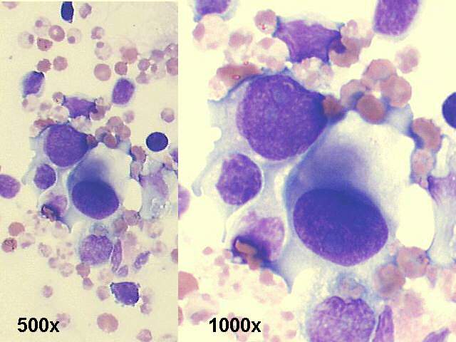
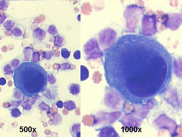
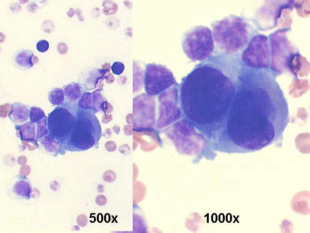
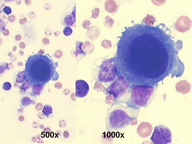
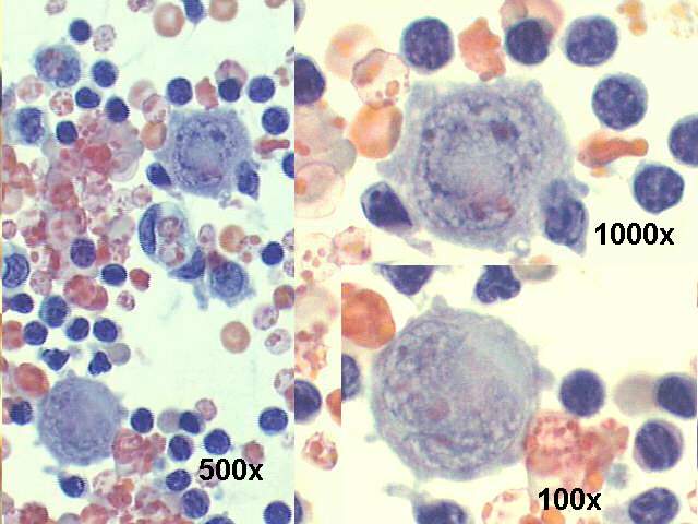
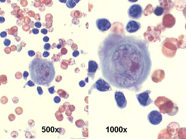
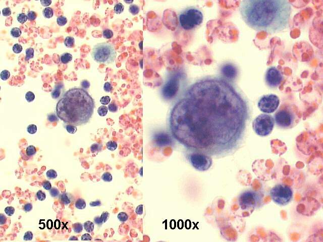
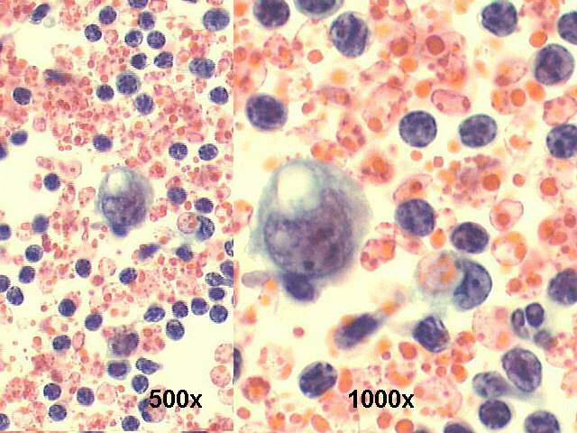
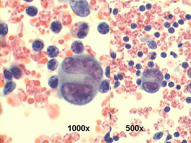
Cytology smears revealed many large isolated (rare groups of two cells were seen) round anaplastic cells, compatible with metastatic lesion, primary angiosarcoma previously resected. Immunocytochemistry was positive for Factor VIII and CD34, confirming vascular (endothelial) histogenesis. They were negative for cytokeratins, actin, chromogranin, and vimentin.
The cytologic features of angiosarcomas have been previously described by Liu and Layfield, in 1999, by Nguyen and Husain, in 2000, and by Boucher et al. in 2000. Boucher et al described vasoformative structures in 10 of their cases, consisting of microacinar structures, arborization of tissue fragments, intracytoplasmic lumens, and signet ring cells. Our case did not demonstrate such structures, except for some signet-ring like cells. In addition, intracytoplasmic hemosiderin was not identified.
References:
Liu K, Layfield LJ. Cytomorphologic features of angiosarcoma of fine needle aspiration biopsy. Acta Cytol 1999;43:407–415.
Boucher L, et al. Cytology of angiosarcoma. Am J Clin Pathol 2000;114:210–219.
Nguyen GK, Husain M: Fine-needle aspiration biopsy cytology of angiosarcoma. Diagn Cytopathol 2000; 23: 143-145.
