Adenocarcinoma metastasis from lung: FNA adrenal mass


Drs. Prolla and Diehl's INTERESTING CASE OF THE MONTH February 2003 case A

Adenocarcinoma metastasis from lung, 200x M-G-G staining

Adenocarcinoma metastasis from lung, 200x M-G-G staining

500x M-G-G, acinar pattern, with peripheral location of the nuclei

Adenocarcinoma metastasis from lung, 500x M-G-G staining, acinar pattern, with peripheral location of the nuclei

Adenocarcinoma metastasis from lung, 500x M-G-G staining, acinar pattern, with peripheral location of the nuclei

Adenocarcinoma metastasis from lung, 500x Pap staining, acinar pattern, with peripheral location of the nuclei

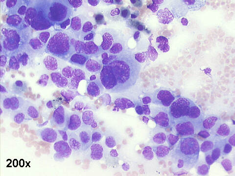
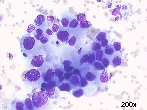
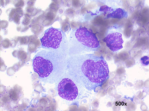
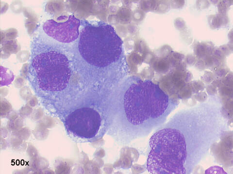
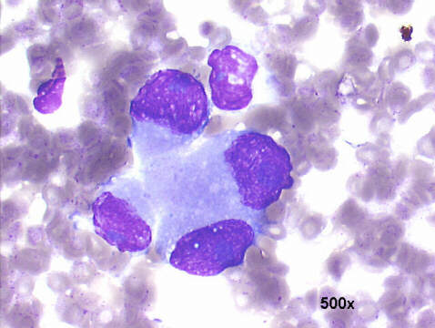
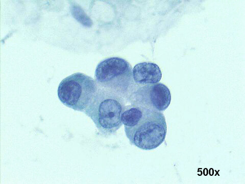
Adenocarcinoma: the smears show loose aggregates of malignant epithelial cells, some with pseudoglandular and acinar patterns, with peripheral location of the nuclei, with coarse chromatin and large nucleoli, typical of adenocarcinoma. The cytological diagnosis was adrenal metastases from adenocarcinoma of the lung. The fact the patient has bilateral adrenal masses also favors this diagnosis of metastatic rather than primary adrenal malignancy.


Case A Feb 2003 Case C Feb 2003 References List of cases Atlas
Hosted by www.Geocities.ws

1