|
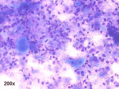
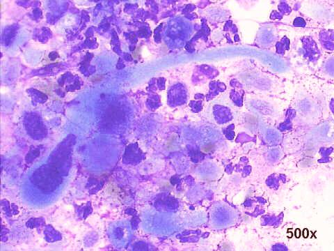
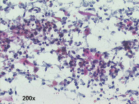
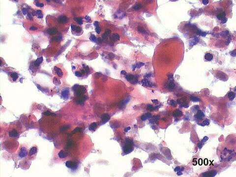
The smears show many fiberlike squamous cells, and others with bizarre shapes (v.g. a tadpole cell), and ghost keratinized cells. There are also many necrotic debris. This is typical of squamous cell carcinoma. A biopsy was diagnostic, and confirmed the cytological findings.
Features of Squamous cell carcinoma
Cellular patterns:
Sheet-like groups of keratinized cells, with distinct cellular borders
Many isolated fiber-like or tadpole cells
Many bizarre-shaped cells
Cytoplasm:
The keratinized cells have orangeophilic/eosinophilic glassy cytoplasm in Papanicolaou staining
In Romanowsky staining they have pale blue or clear cytoplasm
Nucleus
Hyperchromatic, with angular shape
Large nucleoli in non-keratinized cells
Mitoses, karyorrhexis and necrotic debris are frequent
Tumor diathesis |
See also Case B

|