Drs. Prolla and Diehl's INTERESTING CASE OF THE MONTH January 2009
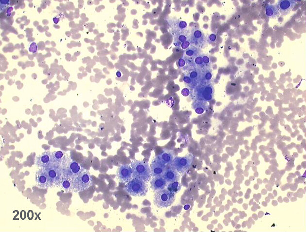
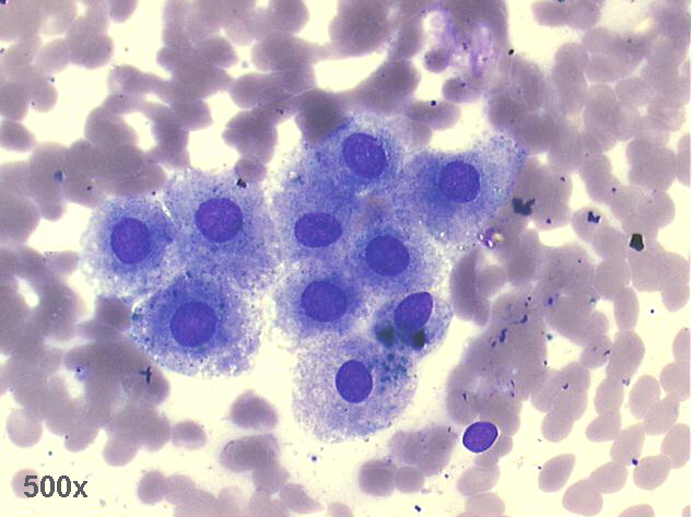
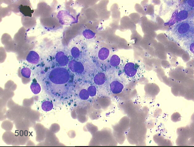
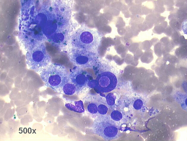
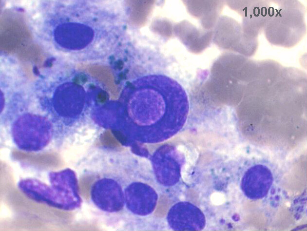
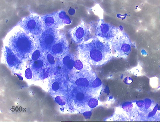
FNA (US guided) Portal vein thrombus, 51-year old female with hepatic tumor:
well differentiated hepatocarcinoma in thrombus.

200x M-G-G staining, groups of hepatocytes

500x of previous image, M-G-G staining, hepatocytes nearly normal looking

500x M-G-G staining, group of hepatocytes with marked anisocytosis and anisonucleosis, obvious bile pigment staining

500x M-G-G staining, group of hepatocytes with marked anisocytosis and anisonucleosis, one large nucleus with intranuclear cytoplasmic inclusion, obvious bile pigment staining

1,000x M-G-G staining, high power view of previous image

500x M-G-G staining, group of hepatocytes with marked anisocytosis and anisonucleosis, less bile pigment staining

The smears show a great number of hepatocytes, with anisocytosis and anisonucleosis, some of them in groups, and others as isolated cells. Most of them have bile pigment in their cytoplasm. In conclusion, the FNA detected that the thrombus was of malignant nature, from a welll differentiated hepatocarcinoma. This interesting procedure has been documented previously: de Sio I. et al: J. Gastroenterol Hepatol 10:662-665, 1995; Dusenbery D, Dodd GD 3rd, Carr BI. : Cancer 75:2057-2062,1995; Lai R, Stephens V, Bardales R.:Gastrointest Endosc. 59:574-577, 2004. For these and other PubMed abstracts see below:


Case January 2009 PubMed Abstracts List of cases Atlas
Hosted by www.Geocities.ws

1