Drs. Prolla and Diehl's
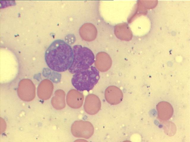
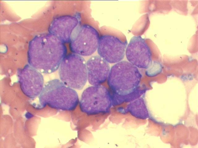
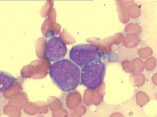
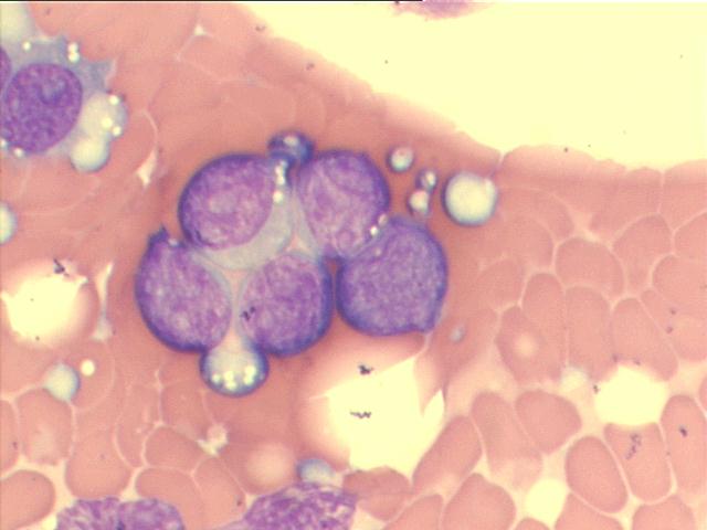
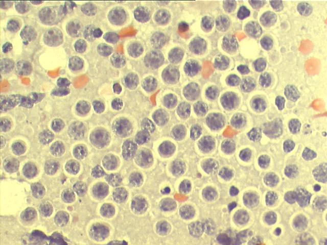
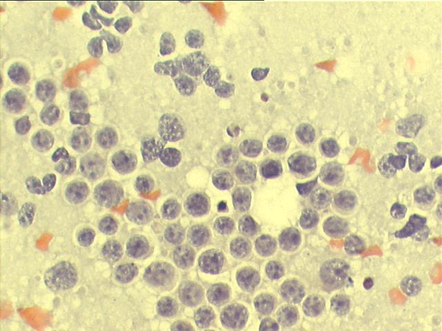
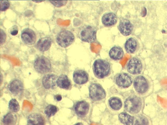
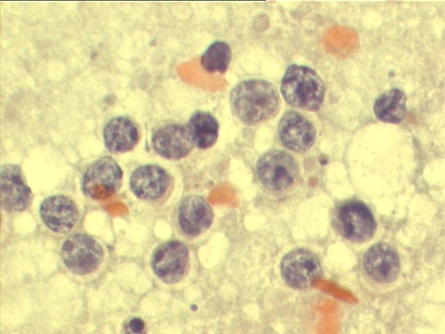
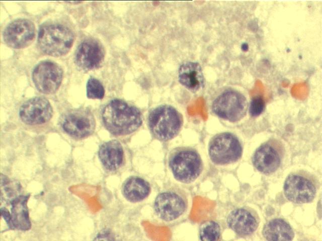
INTERESTING CASE OF THE MONTH January 2008 FNA of breast masses of 28-year old female, with AIDS and B-cell lymphoma
The smears show a great number of large isolated lymphoid cells, with irregular clumped chromatin, suggesting breast involvement by the B-cell lymphoma. Lymphoglandular bodies, pale blue globules of cytoplasmic nature, in cytology smears from fine-needle aspirates, stained by M-G-G or other Romanovsky methods, have long been accepted as being highly suggestive of lymphoid tissue. The lymphoma cells did not show any cohesion, a feature that is seen at least focally in most carcinomas. They also had very little cytoplasm and the nuclei had lymphoid features appropriate to the type of lymphoma, with coarse chromatin and prominent multiple nucleoli. Biopsy of the breast confirmed the presence of high grade B-cell lymphoma in it.