Drs. Prolla and Diehl's INTERESTING CASE OF THE MONTH January 2006
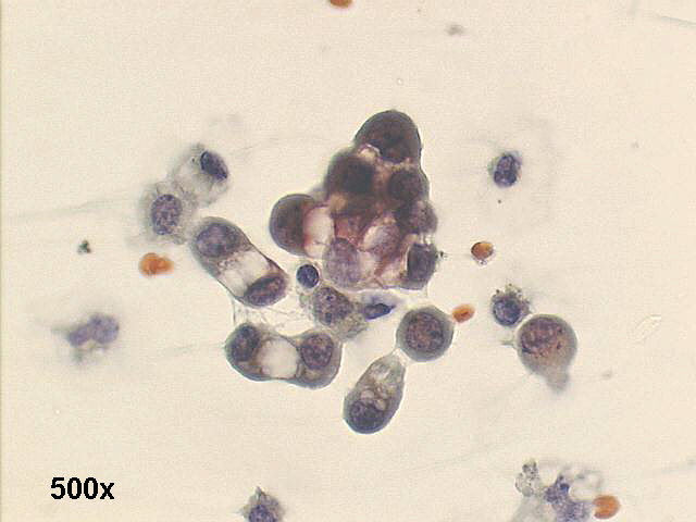
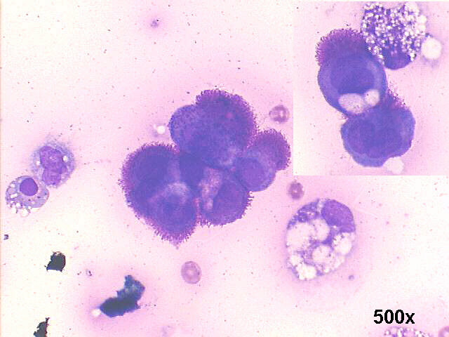
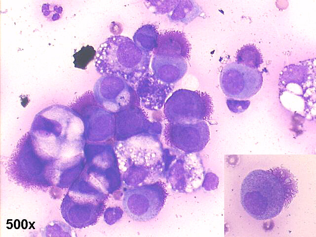
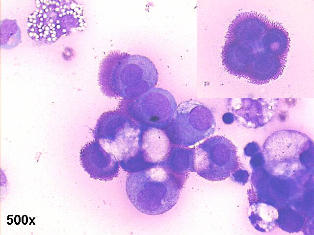
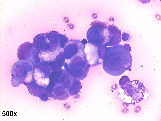
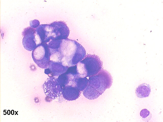
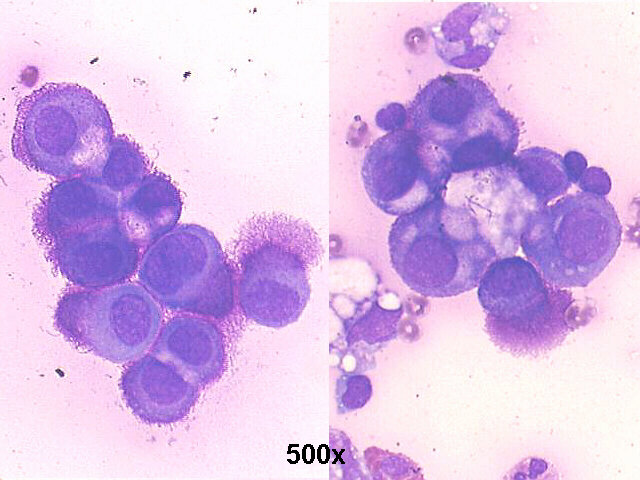
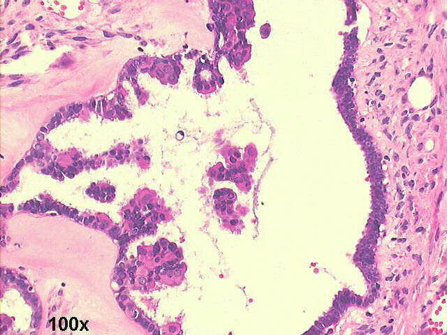
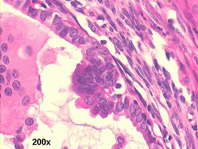
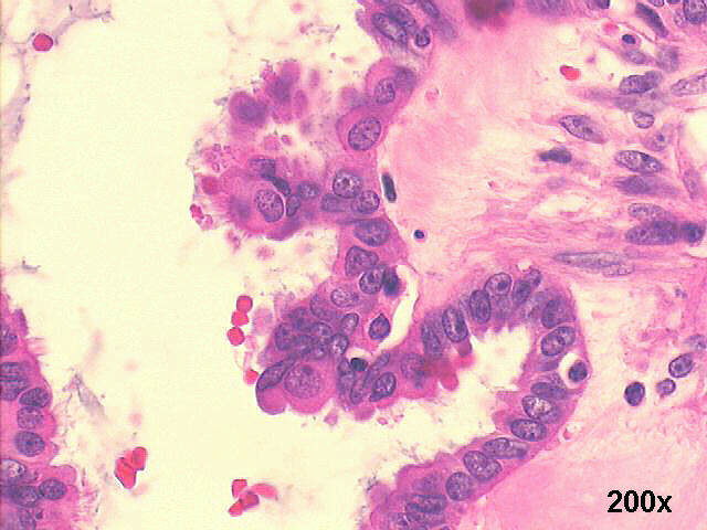
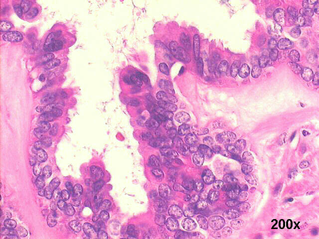
Ascites cytology, 34-year old female:
Papillary carcinoma of ovary. 

Ascites cytopathology: 500x oil Pap staining, very ostensive windows between the cells

Papillary carcinoma of ovary, 500x M-G-G staining, many cells with microvilli, in the inset a window between cells

Papillary carcinoma of ovary, 500x M-G-G staining, many anemone cells with microvilli, some windows between cells

500x M-G-G staining, many cells with microvilli

Papillary carcinoma of ovary, 500x M-G-G staining, many cells with microvilli

Papillary carcinoma of ovary, 500x M-G-G staining, many anemone cells with microvilli

500x M-G-G staining, many anemone cells with microvilli

HISTOLOGY

100x H&E staining, many cells with microvilli

200x H&E staining, many cells with microvilli

200x H&E staining, many cells with microvilli

200x H&E staining, many cells with microvilli

Anemone cells: the presence of many microvilli in malignant cells is frequently documented by electron microscopy (EM), but their presence can be seen in some cases by optical microscopy as well. The use of Romanovsky stains can be very useful in this context ( I use to say they are the EM of the poor...). Spriggs and Boddington, in their classic book Cytology of effusions, Grune & Stratton, 1968, 2nd edition, were the first to call attention to this finding in papillary carcinomas of the ovary, with pictures remarkably similar to the ones in our cases. More recently, this finding has been called "anemone cells" and "anemone tumors" in relation to the cells or the tumors bearing them, respectively.
This case illustrates the difficult differential diagnosis between this type of tumor and mesothelioma, in special of the surface of the ovary and the pelvic peritoneum. Both tumors may exhibit windows between the cells, and microvilli in their surface. There is no immunochemistry test of real value, and the only certainty is done by the surgical exploration, as done in this case, where the pelvic peritoneum was free of disease, and the ovary had a typical serous cystadenocarcinoma papillary tumor. Their coelomic epithelial common origin explains their extreme similarities. Sometimes they both have ciliated cells besides microvilli in their surface.


Back to Case January 2006 References
List of cases
Atlas
Hosted by www.Geocities.ws

1