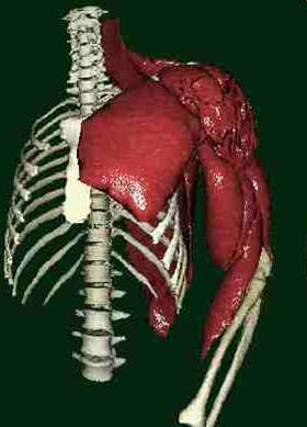
Virtual Reality in Medicine
The CHARM project is a three-years Basic Research Project initiated in November 1993 by the European Commission (EC) under the ESPRIT program. The objective of the European ESPRIT Project CHARM is to develop a Comprehensive Human Animation Resource Model allowing the 3D reconstruction of the human body from medical images and the dynamic simulation of its complex musculoskeletal structure including the simulation of muscular contraction and the finite element deformation of the soft tissues.
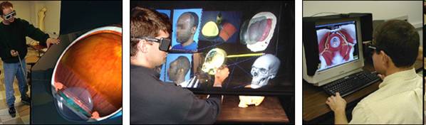
The Georgia Institute of Technology and the Medical College of Georgia have also developed a vr of a proof-of-concept eye surgery simulation that provides both visual and tactile feedback while a surgeon operates on a computer model of the eye in a virtual environment. In practice, ophthalmic surgeons operate on an eye by looking through a stereo microscope while steadying their hands (holding the surgical instruments) on a wrist rest that surrounds the patient's head.
The simulator also includes a stereo operating scope and a wrist rest, but instead of looking directly at a real eye, the surgeon interacts with a virtual eye using a virtual surgical instrument controlled by a hand held 3D position tracking stylus that continuously reports position and orientation to the computer. The tip of the stylus is connected to three motors that generate component force feedback in response to the tool-tissue interaction.
Physicians Personal VR System
The Physician's Personal VR Display is optimized to support consultation from the physician's desk. This display is a conventional PC that supports interaction, stereo, a modest angle-of-view and (optionally) viewer-centered perspective along with teleconferencing. It is meant to facilitate consultation from the physician's desk without requiring that the physician go to a specialized facility. It supports stereo using frame sequential stereo on a conventional CRT PC display and utilizes gain controllers for 3-D interaction.
The simulation includes options to change instruments, record and playback training sessions, reset the models, and peel away outer layers of the eye to reveal interior anatomical components. Dials allow the surgeon to rotate the model, change transparency, zoom, and adjust stereo viewing parameters. An instrument activation switch on the stylus controls actions such as opening and closing forceps and scissors.