
1. Mouth: Including teeth, Oral Cavity, Salvary glands, lips, tougue, and palate; physical digestion; first place that digestion occurs.
2. Pharynx: consists of Epigolottis (flap of cartilage that allows air into trachea and food in esophagus), Larynx (voice box), and Nasal Passage, which is an organ in respiratory system.
3. Esophagus: a long tube that transports food to the stomach.
- Involuntary muscle contraction.
- Paristalsis: Mixes and regulates flow of substances through small intestines; physical digestion; swallows food to the stomach.
- Lower Esophageal Sphineter: funtions in closing of the entrance of stomach, and preventing regargitation.
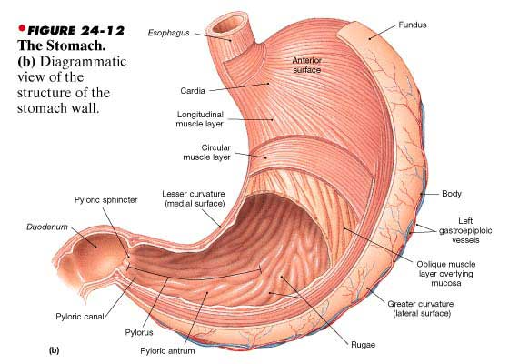
4. Stomach: "J" structure located inferior to diaphagram, and proximal to the liver in abdoninal cavity; Secretes enzyme to break down protein and fats.
- Gastric Glands: Secrete gastric juice to diaphagram; contains pepsin.
- Pepsinogen: Secretes pepsin when it is contracted with HCI.
- Gastrin and Histamine: Promote secretion
- Chyme: mixture of food, mocous and gastric juices; paristalsis contractions mix the chyme
-Gastric Inhibitory Peptide (GIP): a hormone that rebulate the close and open of pyloric sphineter; it is secreted by duodenum in response to acid and fat; provides time for acid to be neutralized and fat digestion in duodenum.
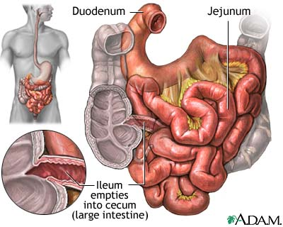
5 Small Intestine: the Longest portion of the body, approximately 5-7 feet; covered by great omentum; the final of digestion, and the aborption of nutrientes through the other body organs.
- Duodenum: Attached to stomach, Common bile duct and pancreas; approximately 25 cm.; posterior to parietal peritoneum; a place where most amount of digestion take place.
- Jejunum: located between duodenum and ileum; where absorptions take place.
- Ileum: the large portion of small intestine; its diameter is lesser than the diameter of jejunum; remainder portionl absorb bable materials, and makes to the ends.
- Mesentary Proper: a layer that supports jejunum and ileum, and contains blood vessels and nerves of lymphatic vessels.
- Villi: Tiny, fingerlike projection that extends outward from the inner lining of small intestine; most in duodenum; contains tiny blood vessels; absorb nutrients and transport them to the body organs.
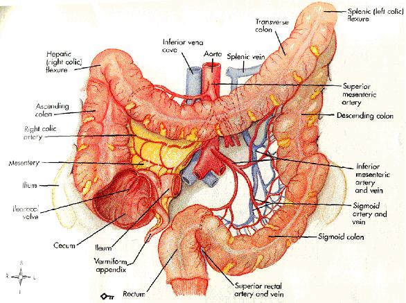
6. Large Intestine: 1.5 meters long; begins at the lower right portion of abdominal cavity; functions in absorption of water, and forms and stores feces. Part of large intestine including:
6.1. Caecum: begining of large intestine; appendix (vestigical organ)
6.2. Colon: divided into four portions: Ascending, Tranverse, descending, and sigmoid colons; absorbing water.
6.3. Rectum: located in the palvic cavity
6.4. Anus: open to outside of the body where feces are expelled; voluntary muscle contraction