 |
| Scientific Interests |
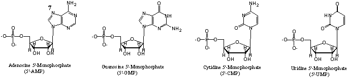
| Interaction of Anti-tumor Ruthenium Complexes with Nucleotides. Many ruthenium complexes show anticancer activity, by inhibiting DNA replication and mutagenic activity, and reducing RNA synthesis [1]. Therefore, finding the interaction site(s) of anti-tumor ruthenium complexes with mononucleotides (5'-AMP, 5'-GMP, 5'-CMP and 5'-UMP, see Fig. 1) is of great importance. |
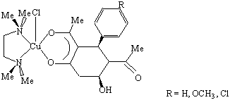
| As a M.Sc project, I selected the anti-tumor active Ru-complexes [RuIII(NH3)5Cl]Cl2, which was known to interact with the N(7) site of purine bases (Fig. 1), and (ImH) trans-[(Im)2Cl4Ru] (Im = imidazol) which showed strong anti-tumor activity [2-3]. By comparing IR spectra of these two complexes using the first complex with known interaction sites as model, we could conclude that the imidazole-Ru complex interacts with 5'-AMP and 5'-GMP in the N(7)-position of the base, and also with an oxygen atom of the phosphate group. |
| Figure 1. Mononucleotides |
| Hydration of Calcium(II) and Uranium(IV). During past few years, my studies have been focused on hydration of ions of biochemical or environmental interest. As a member of Professors M. Sandstr�m and I. Persson group, I took part in an EXAFS study of the hydration of the uranium(IV) ion in aqueous solution, the chemistry of which is of interest when storing nuclear waste in the ground. The number of coordinated water molecules for the U4+ and Th4+ ions was obtained as 10 � 1, the highest hydration number found so far for metal ions. We could also study the complex formation with fluoride ions in solution using EXAFS method [4]. |
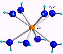
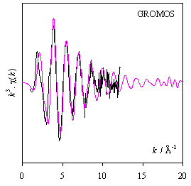
| We also performed a study of the hydration of the calcium ion, a notoriously difficult problem due to the flexibility of its hydration shell in solution, which is also the key to the many important biochemical functions of this ion. For this investigation we had to use several techniques, combining EXAFS with Molecular Dynamics simulations, and calibrate the interaction potentials against results from large angle x-ray scattering. We could conclude that 8 water molecules hydrate the calcium ion in a flexible square-antiprismatic arrangement with an asymmetric distribution of the Ca-O [5]. Both the results and the combination of methods [6] have been received with interest at conferences and meetings [7]. |
| Figure 2. CaCl2 aqueous solution (1 M); experimental EXAFS oscillations compared with theoretical calculated from an MD-simulation. The mean Ca-O distance is 2.46(1) � for an asymmetric distribution of the 8 Ca-O bond distances of the hydrated [Ca(H2O)8]2+ ion |
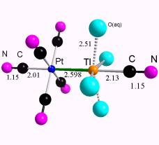
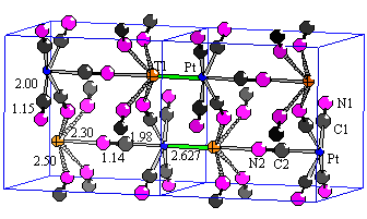
| EXAFS studies on Cyano complexes. The application of EXAFS and vibrational spectroscopy on cyano complexes is another type of structural problem in which I am involved. Technically, EXAFS data from such systems are difficult to interpret due to the excessive multiple scattering within the linearly coordinated cyano ligands. Only recently, the data treatment technique has developed (by ab initio calculations using the programs FEFF (and GNXAS) to a stage where theoretical evaluation of the multiple scattering pathways in such systems has become possible. I could then apply these methods on a number of systems with heterobimetallic platinum-thallium cyano complexes of interest for light-to-energy conversion. For evaluating the structure of a powdered crystalline sample (Fig. 3a), a combination of the results from x-ray powder diffraction and Raman and FT-IR methods proved to be very efficient. In collaboration with Professor J�nos Mink, Veszprem, Hungary, we performed factor group and normal coordinate analyses of the vibrational spectra. We could in this way establish the role of the strongly bridging cyano ligands for the structure and properties of this compound, and evaluate the strength of the Pt-Tl bond, also for a number of heterobimetallic Pt-Tl complexes in aqueous solution (see e.g. Fig. 3b). |
| Figure. 3a. The TlPt(CN)5 compound, two unit cells showing the linear -N2-C2-Pt-Tl-N2-C2-Pt- chains along the c-axis; b. The structure of the [(NC)5Pt-TlCN(aq)]- complex (distances in �). |
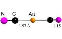
| A number of other cyano complexes, mostly their solution structures, are also under investigation by a combination of EXAFS and vibrational spectroscopy. One purpose is to investigate the character of the M-C bonds. The strong covalency in the Au(I)-cyano complexes is evident from a comparison with the isoelectronic d10 ions Hg(II) and Tl(III) in corresponding linear NC-M-CN complexes. This is shown from the force constants evaluated by normal coordinate analyses but also from the M-C distance, which normally become shorter in such a series when the charge at the central metal atom increases. However, the M-C bond length is found to be for [Au(CN)2] 1.97(1) �, [Hg(CN)2] 2.04(2) � [8], and [Tl(CN)2]+ 2.09(2) � [9]. |
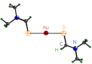
| Similar covalency effects for the Au(I) and Hg(II) ions were found also for the complex formation with a sulfur donor ligand, N,N-dimethylthioformamide. In the crystal structure of [Hg(SCHN(CH3)2](ClO4)2 [10], the Hg-S bond distance of the [Hg(dmtf)2]2+ complex is 2.350(2) �. However, our EXAFS study of the [Au(dmtf)2]+ complex shows a shorter corresponding Au-S bond distance of 2.28(1) �, see Figure 5. Another feature is that Hg(II) readily forms higher complexes both with an excess of cyanide ions and of N,N-dimethylthioformamide, but with these ligands no complexes higher than the linear bis-complexes have been found with gold(I). |
| Figure 4.� The [Au(CN)2] - complex in solution���� |
| (unpublished EXAFS results) |
| Figure 5. Solution structure of the [Au(dmtf)2]+complex; |
| Au-S bond distance: 2.28 �. (unpublished EXAFS results) |
| Thus, the inflexibility of the Au(I) coordination is a special property of its covalency, which is connected to the participation of 6dz2 orbitals in the bonding (a second order Jahn-Teller effect), [10]. This has implications for the biochemical actions of these two metal ions. Mercury(II) is highly toxic, while the biochemistry of gold is mostly connected to the use of gold(I) compounds in treating rheumatoid arthritis. Many drugs are gold(I) thiolates with typically a linear S-Au-S entity with Au-S bond distances of about 2.29 �. Recent findings show that [Au(CN)2]- is a common metabolite of gold(I)-thiolates, and may have a role in their biochemical function [11]. |
| Interesting Demonstrations for Teaching |
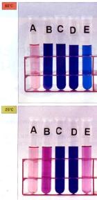
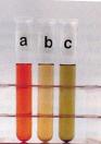
| Solvatochromism - Observation of solvent effects on metal ions. The reversible color change of a compound by changing the external conditions is known as chromotropism. These external conditions can be temperature (thermochromism), pressure (piesochromism), solvent (solvatochromism), or photons with different energies (photochromism). |
| � Solution of CoCl2.6H2O salt is an interesting example. The figure below shows the color of CoCl2 solution in: water (A), methanol (B), ethanol (C), acetone (D) and ammonium chloride aqueous solution (E) at 60 �C (top) and room temperature (below). Its methanol solution shows thermochromic behavior as well [13]. |
| Mixed ligand complexes of copper(II) and nickel(II) with acetylacetone (Hacac) and N,N,N',N'-tetramethylethylenediamine (tmen) show interesting chromotropic properties [14]. [Cu(acac)(tmen)]BPh4 is used as a color indicator for donor properties of solvents [15]. [Ni(acac)(tmen)] BPh4 is red in 1,2-dichloroethane (a), while its methanol solution is green (c). Its solution in acetone (b) shows thermochromic properties. |
| During my Ph.D. studies with Professor Fukuda in Japan, I became interested in solution chemistry, learning about important ligand and solvent properties (such as donor-acceptor, soft-hard properties). I synthesized and characterized a new triketonate ligand and its mixed-ligand copper(II) complexes, which showed solvatochromic behavior in solution and solid states.� The observed absorption maxima (max) of these complexes in various organic solvents show a linear relation versus the acceptor number (AN) of solvent used [16]. |
| References |
| 1) Clarke, M. J., Advances in Chemistry Series, 1997, 253, 349-365. |
| 2) Keppler, B. K.; Rupp, W.; Juhl, U. M.; Endres, H.; Niebel, R. and Bazler, W., Inorg. Chem., 1987, 26, 844-846 & 4366-70. |
| 3) Keppler, B. K., Stenzel, B., Lipponer, K. G., Niebl, R. and Vongerichten; V. E., Noble Met. Biol. Syst. , 1992, 323-48. |
| 4) Moll, H.; Denecke, M. A.; Jalilehvand, F.; Sandstr�m, M. and Grenthe, I., Inorg. Chem., 1999, 38, 1795. |
| 5) Jalilehvand, F.; Sp�ngberg, D.; Lindqvist-Reis, P.; Hermansson, K.; Persson I. and Sandstr�m, M.,� J. Amer. Chem. Soc., 2001, 123, 431. |
| 6) Sp�ngberg, D.; Hermansson, K.; Lindqvist-Reis, P.; Jalilehvand, F.; Sandstr�m M. and Persson, I.,� J. Phys. Chem. B., 2000, 104, 10467. |
| 7) Sandstr�m, M.; Persson, I.; Jalilehvand, F.; Lindqvist-Reis, P.; Sp�ngberg, D. and Hermansson, K., J. Synchr. Rad. 2001, 8, 657. |
| 8) �kesson, R., Persson, I., Sandstr�m, M. and Wahlgren, U., Inorg. Chem.,1994,33, 3715-3723. |
| 9) Jalilehvand, F., Glaser, J., Maliarik, M., Mink, J., Persson, I., Persson, P. Sandstr�m, M. and Toth, I., 2000, in F. Jalilehvand, "Structure of Hydrated Ions and Cyano Complexes", Ph. D. Thesis, KTH, 2000. |
| 10) St�lhandske, C. M. V, St�lhandske, C. I., Sandstr�m, M. and Persson, I., Inorg.Chem., 1997, 36, 3167. |
| 11) Shaw, C. F., in Gold, "Progress in Chemistry, Biochemistry and Technology", Ed. Schmidbaur, H., Wiley, Chichester, 1999, Part II: Biochemistry. |
| 13) F. Jalilehvand and Y. Fukuda, Chemistry Today, Oct. 1994, 51 (Jap.) |
| 14) a) Fukuda, Y., Shimura, A., Mukaida, M., Fujita, E. and Sone, K., J. Inorg. & Nucl. Chem., 1974, 36, 1265; b) Fukuda, Y. and Sone, K., J. Inorg. & Nucl. Chem., 1975, 37, 455. |
| 15) Soukup, R.W. and Schmid, R., J. Chem. Educ., 1985, 62, 459. |
| 16) Jalilehvand, F.; Ishii, Y.; Hidai, M. and Fukuda, Y., J. Chem. Soc., Dalton Trans., 1996, 3251. |
|
 |
 |
 |
 |
 |
 |
 |
 |
| Gems: The Most Beautiful Inorganic Compounds in Nature |
| Research |
 |
| a |
| b |
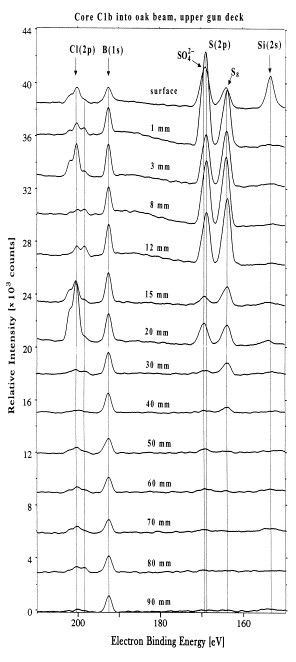
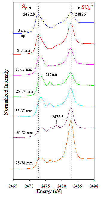
| Sulfur XANES studies on marine-archaeological wooden artefacts. The famous 17th-century Swedish warship Vasa has been on display in the Vasa Museum since 1990. The Vasa was recovered in 1961 after 333 years in the cold brackish water of Stockholm harbor. After extensive conservation treatment the oaken Vasa appeared in good condition. However, high acidity and a rapid spread of sulfate salts were recently observed on many wooden surfaces. We applied X-ray Absorption Spectroscopy (XANES) at the sulfur K-edge recorded in Stanford Linear Accelerator Center (SSRL) for the first time on wooden core samples to speciate the sulfur compounds [12]. The XANES spectra unexpectedly revealed large amounts of the embedded elemental sulfur (0.2-4 mass%) in the wood, also sulfate and in minor amounts several sulfur compounds of intermediate oxidation states (Fig. 8). In humid museum atmospheres, a stepwise sulfur oxidation produces sulfuric acid: S (s) + 3/2 O2 + H2O ----> 2H+(aq) + [SO4]2- X-ray Photoelectron Spectroscopy (XPS) was used to quatify all elements at different depth (Fig. 9). The oxidation is catalyzed by iron species in the wood released from the completely corroded original (8500) iron bolts, as well as from those inserted after salvage. The overall quantity of sulfur in the Vasa is enough to produce more than 5000 kg of sulfuric acid when fully oxidized. Acidic wood hydrolysis is a severe threat to the continued preservation of the Vasa, and pH-raising treatments must be applied to arrest the wood degradation. |
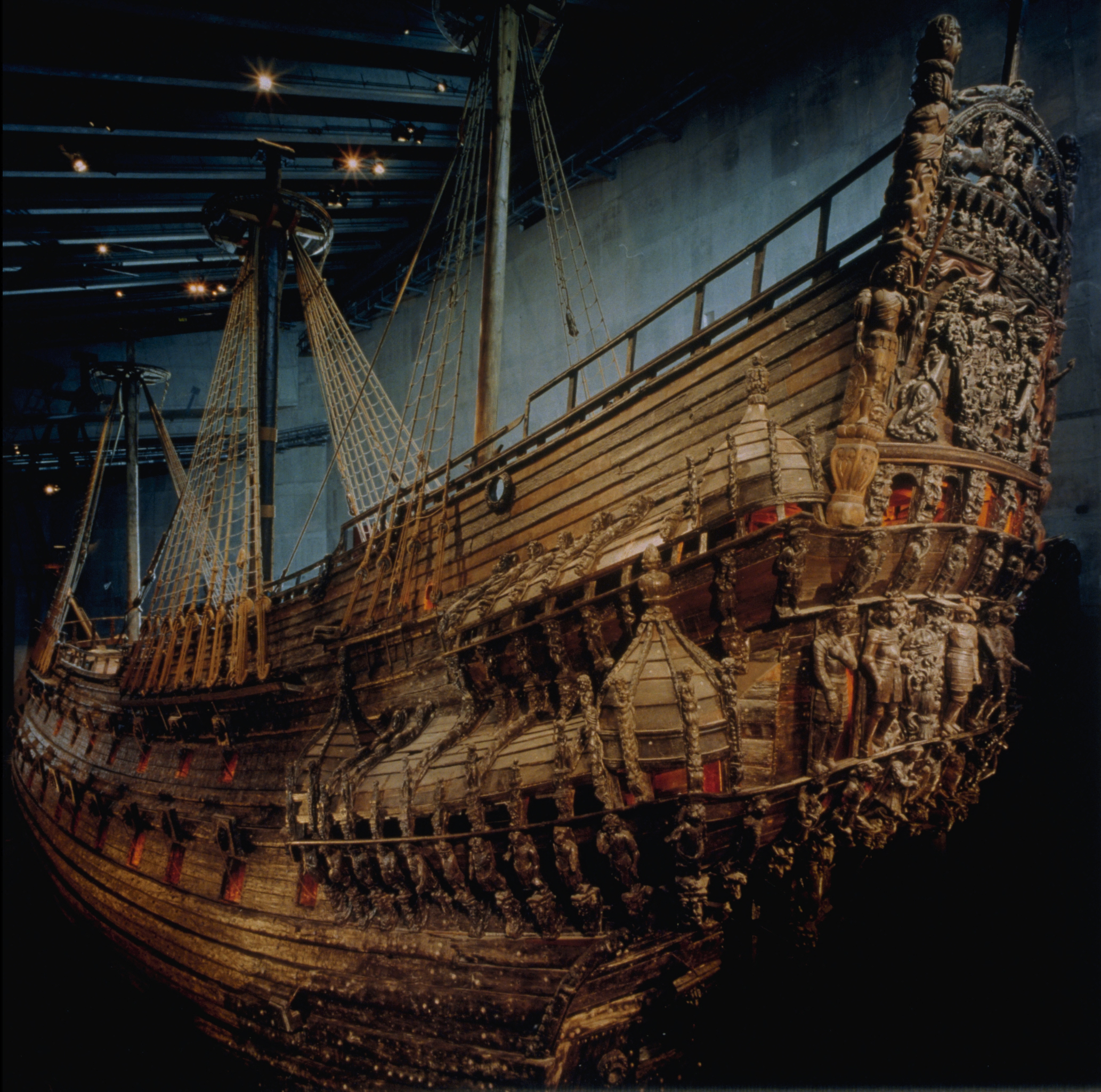 |
| Figure 6. The 17th-century Swedish warship, Vasa (Photo: Hans Hammarskiold / Vasa Museum, Stockholm) |
| But what is the source of the sulfur? The Vasa had sunk to a depth of 32 meters, and the lack of oxygen there inhibited wood-metabolizing microbes. But this environment favors Sulfate Reducing Bacteria that convert sulfate ions in seawater to hydrogen sulfide. During the hundreds of years that Vasa was submerged, hydrogen sulfide penetrated into the deepest layers of the wood. Under the sea, chemical reactions turn hydrogen sulfide into elemental sulfur or pyrite, depending on the amount of the available iron ions. |
 |
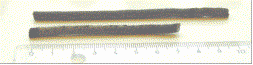
| Figure 7. Vasa Core samples |
 |
 |
| There are similar problem for other wooden marine-archaeological artefacts. Cores from the Dutch Batavia, wrecked outside Western Australia in 1629, were found to contain mostly elemental sulfur and sulfate but also iron sulfides in wood with high iron content. Thus the sulfur problem seems to be general and serious for waterlogged marine-archaeological wood recovered from anoxic conditions. |
| Figure 8. Sulfur K-edge XANES spectra of a core sample of Vasa at different depth, showing the stepwise oxidation of sulfur to sulfuric acid. |
| Figure 9. XPS spectra of a core sample of Vasa, showing the quantity of non-oxidized and oxidized forms of sulfur at different depth. |
| 12) Sandstrom, M.; Jalilehvand, F.; Persson, I.; Gelius, U.; Frank, P. and Hall-Roth, I., Nature., 2002, 415, 893. |