Lymphomatoid
papulosis./Papulosis linfomatoide
Data-Medicos
Dermagic/Express No. 2-(86)
12 Enero 2.000 12 January 2.000
EDITORIAL ESPANOL
=================
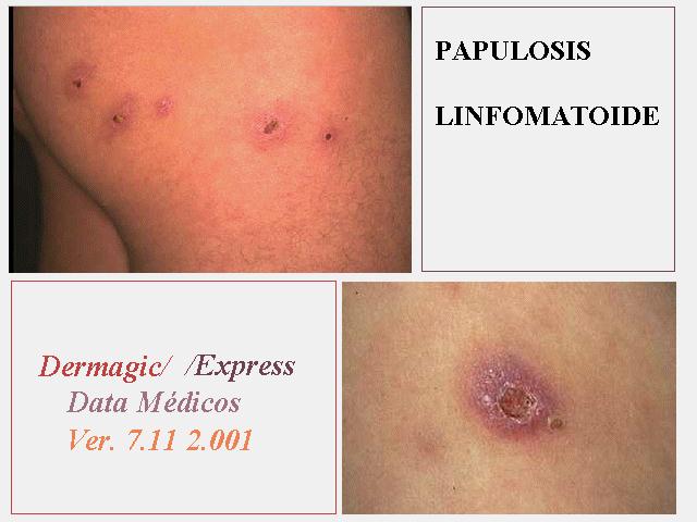
Hola amigos de la red, DERMAGIC de nuevo con ustedes, el tema de hoy
LA
PAPULOSIS LINFOMATOIDE, enfermedad de la piel que bien esta demostrado
es
precursora de malignidad (LIMFOMA). Su relacion con la PITIRIASIS LIQUENOIDE
AGUDA Y CRONICA tambien esta demostrada y bien documentada. El tratamiento,
dificil.
Les puedo asegurar que si examinan bien estas fotos su primer diagnostico
probablemente sea: INFECCION de la piel (IMPETIGO CONTAGIOSO) y su
ultimo:
PAPULOSIS LINFOMATOIDE,,, es todo lo contrario... se trata de una PAPULOSIS
LINFOMATOIDE. Espero disfruten estas 65 referencias.
Les recuerdo que dermagic sera quincenal a partir de esta fecha ,,,
Saludos a todos !!!
Dr. Jose Lapenta R.,,,
EDITORIAL ENGLISH
=================
Hello friends of the net, DERMAGIC again with you, today's topic THE
LYMPHOMATOID PAPULOSIS , illness of the skin that is well demonstrated
is
precursor of malignancy (LYMPHOMA). Their relationship with the PITYRIASIS
LICHENOIDES ACUTE AND CHRONIC also is demonstrated and well documented.
The treatment, difficult.
I can assure that if you examine these pictures well your first I diagnose
be probably:
INFECTION of the skin (IMPETIGO CONTAGIOSA) and their I last:
LYMPHOMATOID PAPULOSIS, it is just the opposite... it is a LYMPHOMATOID
PAPULOSIS I wait enjoyments these 65 references.
I remind that dermagic will be biweekly starting from this date,
Greetings to ALL, !!
Dr. Jose Lapenta R.,,,
===================================================================
REFERENCIAS BIBLIOGRAFICAS / BIBLIOGRAPHICAL REFERENCES
===================================================================
=================================================================
1.) Lymphomatoid papulosis associated with pregnancy.
2.) Lymphomatoid papulosis: successful weekly pulse superpotent topical
corticosteroid therapy in three pediatric patients.
3.) Lymphomatoid papulosis and cutaneous CD30+ lymphoma. 4.) Amplification
of
genomic DNA demonstrates the presence of the t(2;5) (p23;q35) in anaplastic
large cell
lymphoma, but not in other non-Hodgkin's lymphomas, Hodgkin's disease,
or
lymphomatoid papulosis [see comments]
5.) A case of lymphomatoid papulosis occurred simultaneously with
Ki-1-positive
anaplastic large cell lymphoma.
6.) Lymphomatoid papulosis type A: clinical, morphologic, and
immunophenotypic study.
7.) Increased risk of lymphoid and nonlymphoid malignancies in patients
with
lymphomatoid papulosis.
8.) Lymphomatoid papulosis in association with mycosis fungoides: a
study
of 15 cases.
9.) Involvement of the tongue by lymphomatoid papulosis.
10.) Lymphomatoid papulosis treated with extracorporeal photochemotherapy.
11.) Absence of anaplastic lymphoma kinase (ALK) and Epstein-Barr virus
gene products in primary cutaneous anaplastic large cell lymphoma and
lymphomatoid
papulosis.
12.) Absence of Epstein-Barr virus in lymphomatoid papulosis. An
immunohistochemical
and in situ hybridization study [see comments]
13.) Methotrexate is effective therapy for lymphomatoid papulosis and
other
primary
cutaneous CD30-positive lymphoproliferative disorders.
14.) Follicular lymphomatoid papulosis.
15.)Is it lymphoma or lymphomatoid papulosis?
16.) Lymphomatoid papulosis: a low-grade T-cell lymphoma?
17.) The dominant T cell clone is present in multiple regressing skin
lesions and associated
T cell lymphomas of patients with lymphomatoid papulosis.
18.) The cytokine mRNA expression in primary cutaneous CD30-positive
lymphoproliferative disorders: successful treatment with recombinant
interferon-gamma.
19.) Low incidence of Epstein-Barr virus presence in primary cutaneous
T-cell
lymphoproliferations.
20.) Clonal disease in extracutaneous compartments in cutaneous T-cell
lymphomas. A comparative study between cutaneous T-cell lymphomas and
pseudo
lymphomas.
21.) Apoptotic and proliferating cells in cutaneous lymphoproliferative
diseases.
22.) No evidence of HTLV-I proviral integration in lymphoproliferative
disorders
associated with cutaneous T-cell lymphoma.
23.) Interleukin-7 receptor expression in cutaneous T-cell lymphomas.
24.) The t(2;5) in human lymphomas.
25.) Pityriasis lichenoides in children: clinicopathologic review of
22
patients.
26.) Regional lymphomatoid papulosis: a report of four cases.
27.) Infection with parapoxvirus induces CD30-positive cutaneous
infiltrates in humans.
28.) CD30-CD30 ligand interaction in primary cutaneous CD30(+) T-cell
lymphomas: A
clue to the pathophysiology of clinical regression.
29.) Increased risk of lymphoid and nonlymphoid malignancies in patients
with
lymphomatoid papulosis.
30.) Primary cutaneous CD8-positive epidermotropic cytotoxic T cell
lymphomas. A
distinct clinicopathological entity with an aggressive
clinical behavior.
31.) Lymphomatoid papulosis in association with mycosis fungoides:
a study
of 15 cases.
32.) Early detection of cutaneous lymphoma.
33.) Cutaneous pseudolymphomas.
34.) Lymphomatoid papulosis in childhood with exclusive acral involvement.
35.) Absence of HTLV-1 proviral sequences in patients with lymphomatoid
papulosis
36.) Clinicopathologic manifestations of Epstein-Barr virus-associated
cutaneous
lymphoproliferative disorders.
37.) Follicular lymphomatoid papulosis and multiple myeloma.
38.) Lymphomatoid papulosis--treatment with recombinant interferon
alfa-2a
and
etretinate.
39.) Lymphomatoid papulosis: a follow-up study of 41 patients.
40.) Lymphomatoid papulosis associated with acquired ichthyosis.
41.) Primary anaplastic large-cell lymphoma of the skin. A case report
suggesting that
regressing atypical histiocytosis and lymphomatoid papulosis are subsets.
42.) Lymphomatoid papulosis: a clinical and histopathologic review
of 53
cases with
leukocyte immunophenotyping, DNA flow cytometry, and T-cell receptor
gene
rearrangement studies.
43.) [Lymphomatoid papulosis and anaplastic giant-cell lymphoma]
44.) Molecular evidence for a clonal relationship between lymphomatoid
papulosis and
Ki-1 positive large cell anaplastic lymphoma.
45.) Lymphomatoid papulosis followed by Hodgkin's lymphoma. Differential
response to
therapy [see comments]
46.) Epidemiology of lymphomatoid papulosis [see comments]
47.) Lethal midline granuloma (peripheral T-cell lymphoma) after lymphomatoid
papulosis.
48.) The prognosis of patients with lymphomatoid papulosis associated
with
malignant
lymphomas.
49.) Immunophenotypic and genotypic characterization of lymphomatoid
papulosis.
50.) Pityriasis lichenoides and lymphomatoid papulosis.
51.) Lymphomatoid papulosis: clinicopathological comparative study
with
pityriasis
lichenoides et varioliformis acuta.
52.) PUVA-induced lymphomatoid papulosis in a patient with mycosis
fungoides.
53.) Accuracy in diagnosis of lymphomatoid papulosis.
54.)Regressing atypical histiocytosis and lymphomatoid papulosis: variants
of the same
disorder?
55.) Lymphomatoid papulosis and its relationship to "idiopathic"
hypereosinophilic
syndrome.
56.) Lymphomatoid papulosis/pityriasis lichenoides in two children.
57.) [Acyclovir in mycosis fungoides and lymphomatoid papulosis]
58.) Eosinophilic histiocytosis: a variant form of lymphomatoid papulosis
or a disease sui
generis?
59.) Lymphomatoid papulosis: a premalignant T cell disorder.
60.) Clinical and histologic differentiation between lymphomatoid papulosis
and pityriasis
lichenoides.
61.) The clinicopathologic spectrum of lymphomatoid papulosis: study
of 31
cases.
62.) [Lymphomatoid papulosis in a child]
63.)Lymphomatoid papulosis in an 11-month-old infant.
64.) Topical carmustine therapy for lymphomatoid papulosis.
65.)Immunohistology of pityriasis lichenoides et varioliformis acuta
and
pityriasis
lichenoides chronica. Evidence for their interrelationship with
lymphomatoid papulosis.
============================================================
============================================================
1.) Lymphomatoid papulosis associated with pregnancy.
============================================================
Author
Yamamoto O; Tajiri M; Asahi M
Address
Department of Dermatology and Occupational Dermatopathology, University
of
Occupational and Environmental Health, Kitakyushu, Japan.
Source
Clin Exp Dermatol, 22(3):141-3 1997 May
Abstract
We report a case of lymphomatoid papulosis which developed in a 29-year-old
pregnant woman. She had numerous papules scattered over the inner aspect
of
the left thigh. Histology of the biopsy specimen demonstrated an atypical
mononuclear cell infiltration of the dermis. Spontaneous regression
of the
lesions occurred after termination of gestation. A possible effect
of
hormonal changes and alterations in T lymphocyte activity during pregnancy
on the occurrence of lymphomatoid papulosis is discussed. In 1968,
Macaulay
introduced the term lymphomatoid papulosis for a chronic self-healing
skin
lesion which was clinically benign and histologically malignant.
Clinically, lymphomatoid papulosis consists of involuting and recurring
papules, plaques and nodules. Histopathologically, the lesion is
characterized by an atypical lymphoid infiltrate which resembles malignant
lymphoma. Immunohistochemically, the atypical lymphoid cells bear T-cell
markers and are characterized by the expression of Ki-1 or CD30. We
describe the first case of typical lymphomatoid papulosis which developed
during pregnancy.
============================================================
2.) Lymphomatoid papulosis: successful weekly pulse superpotent topical
corticosteroid therapy in three pediatric patients.
============================================================
Author
Paul MA; Krowchuk DP; Hitchcock MG; Jorizzo JL
Address
Department of Dermatology, Bowman Gray School of Medicine, Wake Forest
University, Winston-Salem, NC 27157, USA.
Source
Pediatr Dermatol, 13(6):501-6 1996 Nov-Dec
Abstract
Lymphomatoid papulosis is a T-cell proliferation that occurs primarily
in
adults but has been well described in children. Lesions may regress
spontaneously but often leave residual scarring and, as a result,
intervention frequently is considered. Therapeutic modalities commonly
employed for adults with lymphomatoid papulosis may be poorly tolerated
by
pediatric patients. We present a series of three children with lymphomatoid
papulosis treated with superpotent topical corticosteroids (halobetasol
or
clobetasol propionate). When applied twice daily for 2 to 3 weeks followed
by weekly pulsed application, this treatment resulted in complete
resolution of nearly all cutaneous lesions. Three ulcerated lesions,
occurring in two patients, required adjuvant therapy with intralesional
triamcinolone. To date one patient remains free of cutaneous disease
and
two children experience occasional new lesions that respond to renewed
treatment with topical clobetasol propionate. None of the children
have
evidence of systemic disease. We conclude that pulsed application of
a
superpotent topical corticosteroid is efficacious and safe in the
management of cutaneous lesions of lymphomatoid papulosis and avoids
the
risks often associated with more aggressive interventions. Since these
agents do not alter the risk of subsequent malignancy, careful ongoing
surveillance of children with lymphomatoid papulosis is imperative.
============================================================
3.) Lymphomatoid papulosis and cutaneous CD30+ lymphoma.
============================================================
Author
LeBoit PE
Address
Department of Pathology, University of California, San Francisco
94143-0506, USA.
Source
Am J Dermatopathol, 18(3):221-35 1996 Jun
Abstract
Lymphomatoid papulosis and cutaneous CD30+ lymphoma are closely related
conditions in which large atypical lymphocytes that have similar
immunophenotypic features occur. In lymphomatoid papulosis, the lesions
are
papules and nodules that spontaneously involute. There are two polar
histologic patterns, type A and B, in which the large atypical cells
resemble those of Hodgkin's disease and mycosis fungoides, respectively,
but in many cases, features of both types are present, either separately
or
in the same lesions. Variants of lymphomatoid papulosis include cases
with
a perifollicular distribution and those with lymphocytic vasculitis
or
dermal mucin deposits. Clinical lesions that tend to be stable, a
monomorphous cellular composition, and in the case of immunocompromised
patients, the presence of Epstein-Barr viral genome characterize cutaneous
CD30+ lymphoma. A loss of response to transforming growth factor-beta,
which normally dampens cellular proliferation, may differentiate CD30+
lymphoma from lymphomatoid papulosis.
============================================================
4.) Amplification of genomic DNA demonstrates the presence of the t(2;5)
(p23;q35) in anaplastic large cell lymphoma, but not in other non-Hodgkin's
lymphomas, Hodgkin's disease, or lymphomatoid papulosis [see comments]
============================================================
Author
Sarris AH; Luthra R; Papadimitracopoulou V; Waasdorp M; Dimopoulos
MA;
McBride JA; Cabanillas F; Duvic M; Deisseroth A; Morris SW; Pugh WC
Address
Department of Hematology, University of Texas M.D. Anderson Cancer
Center,
Houston 77030, USA.
Source
Blood, 88(5):1771-9 1996 Sep 1
Abstract
Anaplastic large cell lymphoma (ALCL) is a distinct clinicopathologic
variant of intermediate grade non-Hodgkin's lymphomas (NHL) composed
of
large pleomorphic cells that usually express the CD30 antigen and
interleukin (IL)-2 receptors, and is characterized by frequent cutaneous
and extranodal involvement. With variable frequency ALCL bear the
t(2;5)(p23;q35) chromosomal translocation that fuses the nucleophosmin
(NPM) gene on chromosome 5q35 to a novel protein kinase gene, Anaplastic
Lymphoma Kinase (ALK), on chromosome 2p23. We determined the frequency
of
this translocation with a novel DNA polymerase chain reaction (PCR)
technique using 0.5 microgram of genomic DNA, 5'-primers derived from
the
NPM gene and 3'-primers derived from the ALK gene and hybridization
with
internal probes. The presence of amplifiable DNA in the samples was
tested
with the inclusion in the PCR reaction of oligonucleotide primers designed
to amplify a 3016-bp fragment from the beta-globin locus. NMP-ALK fusion
amplicons were detected using DNA isolated either from all three ALCL
cell
lines tested, or from all four primary ALCL tumors known to contain
the
t(2;5)(p23;q35) translocation. Nested amplicons were detected by
hybridization in 100% of specimens diluted 10(4)-fold and in 20% of
those
diluted 10(5)-fold. We subsequently examined archival genomic DNA from
20
patients with ALCL, 39 with diffuse large cell, 2 with mantle cell,
20 with
peripheral T cell, 13 with low-grade NHL, 31 with Hodgkin's disease
(HD),
and 6 with lymphomatoid papulosis. Fusion of the NPM and ALK genes
was
detected in three of 18 patients with ALCL who had amplifiable DNA
(17%,
95% confidence intervals 4% to 41%), but not in any patients with other
NHL, HD, or lymphomatoid papulosis. The amplicon sizes were different
in
all cell lines and patients reflecting unique genomic DNA breakpoints.
We
conclude that with genomic DNA-PCR the rearrangement of the NPM and
ALK
loci is restricted to patients with ALCL. Further studies are needed
to
determine the prognostic significance of the NPM-ALK rearrangement,
to
determine whether its detection can aid in the differential diagnosis
between ALCL. Hodgkin's disease, and lymphomatoid papulosis, and to
establish the usefulness of the genomic DNA PCR in the monitoring of
minimal residual disease in those patients whose tumors bear the t(2;5).
============================================================
5.) A case of lymphomatoid papulosis occurred simultaneously with
Ki-1-positive anaplastic large cell lymphoma.
============================================================
Author
Lee NS; Cha SW; Hong SJ; Shin WY; Lee GT; Jeon JW; Won JH; Baick SH;
Hong
DS; Park HS
Address
Department of Internal Medicine, Soonchunhyang University, College
of
Medicine, Seoul, Korea.
Source
Korean J Intern Med, 12(1):84-8 1997 Jan
Abstract
Lymphomatoid papulosis (LyP) is a chronic self-healing skin eruption
that
is clinically benign but histologically mimics a malignant lymphoma.
However, lymphomatoid papulosis with anaplastic large cell lymphoma
responds poorly to medical treatments, including chemotherapies. We
experienced a 60-year-old male patient with lymphomatoid papulosis
occurred
simultaneously with relapsed Ki-1-positive anaplastic large cell lymphoma
who was treated with salvage chemotherapy but, unfortunately, failed
to be
rescued. We report it with a review of the literature.
============================================================
6.) Lymphomatoid papulosis type A: clinical, morphologic, and
immunophenotypic study.
============================================================
Author
Sioutos N; Asvesti C; Sivridis E; Aygerinou G; Tsega A; Zakopoulou
N;
Zographakis I
Address
Department of Pathology, Georgetown University, Washington, DC, USA.
Source
Int J Dermatol, 36(7):514-7 1997 Jul
Abstract
BACKGROUND: Lymphomatoid papulosis (LyP) is a cutaneous clonal or
polyclonal Ki-1 + T-cell lymphoproliferative disorder, morphologically
resembling Ki-1 + anaplastic large cell lymphomas (Ki-1 + ALCL) or
Hodgkin's disease (HD). Lymphomatoid papulosis usually has a characteristic
benign clinical course with remissions and relapses of the cutaneous
eruptions. METHODS: The authors studied three patients with LyP. In
each
case the diagnosis was established based on the typical clinical history
and presentation of the cutaneous lesions as well as the morphologic
and
immunophenotypic findings. RESULTS: In all three cases the skin biopsies
showed a polymorphic, nonepidermotropic, dermal lymphocytic infiltrate,
composed of small lymphocytes and fewer large, atypical cells. The
large
cells were positive for the activation markers CD30 (Ki-1) and CD45R
(leukocyte common antigen), and were negative for the HD marker CD15
(Leu
MI). CONCLUSIONS: In most cases, LyP can be distinguished from Ki-1
+ ALCL
and HD on the basis of clinical, morphologic, and/or immunophenotypic
findings. We emphasize the importance of the recognition of LyP as
a
clinicopathologic entity and the awareness of dermatologists, oncologists,
and surgical pathologists in differentiating LyP from other primary
cutaneous Ki-1 + lymphoproliferative disorders (Ki-1 + ALCL and HD).
The
prognosis of cutaneous Ki-1 + ALCL and HD is usually different from
LyP and
requires a different therapeutic approach.
============================================================
7.) Increased risk of lymphoid and nonlymphoid malignancies in patients
with lymphomatoid papulosis.
============================================================
Author
Wang HH; Myers T; Lach LJ; Hsieh CC; Kadin ME
Address
Department of Pathology, Beth Israel Deaconess Medical Center and Harvard
Medical School, Boston, Massachusetts 02115, USA.
Source
Cancer, 86(7):1240-5 1999 Oct 1
Abstract
BACKGROUND: Lymphomatoid papulosis (LyP) is a rare skin disease with
malignant potential. The long term outcomes of patients with this disease
have not been adequately assessed. METHODS: Fifty-seven patients with
biopsy-proven LyP and 67 controls matched for age, gender, and race
were
followed prospectively from 1988 to 1996. Reported malignancies were
confirmed by surgical pathology and/or autopsy reports. A search through
the National Death Index through December 1995 was conducted to identify
all deaths, and death certificates were procured. Expected numbers
of
malignancies based on SEER data were calculated for both the patient
and
the control groups. RESULTS: Six LyP patients (10.5%) and 1 control
(1.5%)
reported nonlymphoid malignancies (P = 0.047). Two patients and no
controls
developed lymphoid malignancies (mycosis fungoides and CD30(+) cutaneous
lymphoma). The expected numbers of nonlymphoid and lymphoid malignancies
in
the LyP patient group, based on the SEER data, were 1.93 and 0.15,
respectively, yielding a relative risk (with 95% confidence interval)
of
3.11 (1.26-6.47) for nonlymphoid malignancies and 13.33 (2.24-44.05)
for
malignant lymphomas in the LyP patients. There was no significant
difference between the observed and expected numbers of malignancies
in the
control group. Four LyP patients died during the follow-up, three due
to
malignancies; and one control died of a gunshot wound to the head
(suicide). The difference in overall survival between the LyP patients
and
the controls was not statistically significant (P = 0. 12). CONCLUSIONS:
Patients with LyP appear to have an increased risk of both lymphoid
and
nonlymphoid malignancies. The increased risk of nonlymphoid as well
as
lymphoid malignancies may suggest a basic underlying genetic defect
leading
to the development of malignancy in LyP patients. Copyright 1999 American
Cancer Society.
============================================================
8.) Lymphomatoid papulosis in association with mycosis fungoides: a
study
of 15 cases.
============================================================
Author
Basarab T; Fraser-Andrews EA; Orchard G; Whittaker S; Russel-Jones
R
Address
St John's Institute of Dermatology, St Thomas' Hospital, London, UK.
Source
Br J Dermatol, 139(4):630-8 1998 Oct
Abstract
We report clinical findings in 15 patients with lymphomatoid papulosis
(LyP) associated with mycosis fungoides (MF). LyP either preceded (n
= 4),
followed (n = 5) or occurred concurrently with the MF lesions (n =
6).
Twenty-eight LyP lesions were classified histologically and analysed
further with immunostaining for CD3 and CD30. Five biopsies contained
a
predominance of type A cells, six biopsies contained a predominance
of type
B cells. and six were mixed (A + B). However, 11 biopsies contained
a
population of atypical mononuclear cells with large hyperchromatic
nuclei
that we have termed indeterminate cells. These cells contained a thin
rim
of eosinophilic cytoplasm and showed strong CD30 but absent, faint
or
normal CD3 staining. In seven biopsies from five separate patients
these
cells represented the predominant cell type and we have termed this
the
pleomorphic variant of LyP. Analysis of T-cell receptor genes using
Southern blot analysis and polymerase chain reaction/single strand
conformational polymorphism analysis identified a T-cell clone in six
of 16
LyP lesions and nine of 16 MF lesions. In the three patients who had
clones
in both types of skin lesions, the clones were identical. Only two
of 10
blood samples, both of which were from the same patient, had a T-cell
clone
and none of two lymph nodes showed evidence of a clonal population.
To date
all patients are alive with a median follow-up of 15 years from the
onset
of the first lesion. One patient has developed Lin anaplastic large
cell
lymphoma of the nasopharynx. These data augment the current literature
on
the association of LyP and MF and suggest that the
============================================================
9.) Involvement of the tongue by lymphomatoid papulosis.
============================================================
Author
Kato N; Tomita Y; Yoshida K; Hisai H
Address
Department of Dermatology and Research Institute, National Sapporo
Hospital, Japan.
Source
Am J Dermatopathol, 20(5):522-6 1998 Oct
Abstract
We report on a case of lymphomatoid papulosis (LyP) with involvement
of the
tongue. The patient was a 34-year-old Japanese man. Three reddish,
centrally depressed, slightly elevated nodules were evident on the
dorsal
tongue, along with lesions elsewhere on the skin. One of them was biopsied
and exhibited a superficial and deep, perivascular and interstitial
mixed
cellular infiltrate including atypical lymphoid cells, lymphocytes,
neutrophils, and histiocytes. The patient also showed rhythmical recurrence
of reddish papules and ulcerated nodules on the trunk, extremities,
and
anogenital area. Histologically, these papules showed a dense, wedge-shaped
mixed cellular infiltrate in the dermis, which included medium and
large
atypical lymphoid cells, lymphocytes, neutrophils, and histiocytes.
Immunoperoxidase staining for CD30 was positive in the cell membrane
and
cytoplasm of the atypical cells. We could not find other reports of
LyP
involving the tongue. Systemic treatment with interferon (INF)-alpha2a
was
dramatically effective in inhibiting recurrence of the eruption.
============================================================
10.) Lymphomatoid papulosis treated with extracorporeal photochemotherapy.
============================================================
Author
Wollina U
Address
Department of Dermatology, University of Jena, 07740 Jena, Germany.
Source
Oncol Rep, 5(1):57-9 1998 Jan-Feb
Abstract
Lymphomatoid papulosis (LP) is a rare low-grade T-cell lymphoma which
may
respond to cytotoxic drugs and PUVA irradiation but long-term remission
has
not been achieved. Extracorporeal photochemotherapy (ECP) is an
immunomodulating therapy used successfully for several types of CTCL,
no
experience with LP has been reported yet. ECP therapy with
8-methoxypsoralen was introduced on two subsequent days once per month
for
half a year in a 42-year old women with a 20-year history of LP.
Disseminated papules disappeared rapidly after 3 cycles of ECP treatment
but metastatic spread continued, which made necessary a subsequent
polychemotherapy. One year later, the patient died of central nervous
system metastasis. ECP monotherapy seems unable to control disease
progression in LP despite beneficial effects on skin lesions.
============================================================
11.) Absence of anaplastic lymphoma kinase (ALK) and Epstein-Barr virus
gene products in primary cutaneous anaplastic large cell lymphoma and
lymphomatoid papulosis.
============================================================
Author
Herbst H; Sander C; Tronnier M; Kutzner H; H¨ugel H; Kaudewitz
P
Address
Institut f¨ur Pathologie, Universit¨atsklinikum
Benjamin Franklin, Freie
Universit¨at, Berlin, Germany.
Source
Br J Dermatol, 137(5):680-6 1997 Nov
Abstract
The prevalence of the t(2;5)(p23;q35) and/or anaplastic lymphoma kinase
(ALK) gene products in cutaneous anaplastic large cell (ALC) lymphomas
and
a potential precursor lesion, lymphomatoid papulosis (LyP), is
controversial. ALK gene products, which are absent from normal
lymphohaematopoietic cells, are a phenotypic marker of lymphomas carrying
the t(2;5). We used in situ hybridization and immunohistology to screen
14
cutaneous ALC lymphomas, 21 cases of LyP, and one nodal ALC lymphoma
associated with LyP for ALK gene products. ALK gene products were not
detectable in these cases. In contrast, ALK gene products were found
in a
lymphonodal ALC lymphoma with subsequent extension to the skin and
in
t(2;5)-positive cell lines. Detection of the Epstein-Barr virus
(EBV)-encoded small nuclear transcripts (EBER), and of immunoglobulin
light
chain transcripts served to check for the presence of cellular RNA
in the
tissue sections. EBER transcripts were found in scattered reactive
lymphoid
cells, but not in atypical or tumour cells. ALK gene expression and
EBV
infection seem to be a rare finding in cutaneous ALC lymphomas and
LyP.
This points to a molecular aetiology of primary cutaneous ALC lymphomas
and
LyP distinct from that of extracutaneous CD30+ lymphoproliferative
disease.
Detection of the t(2;5) or ALK gene products in cutaneous
lymphoproliferative lesions therefore requires exclusion of extracutaneous
ALC lymphoma in such patients.
============================================================
12.) Absence of Epstein-Barr virus in lymphomatoid papulosis. An
immunohistochemical and in situ hybridization study [see comments]
============================================================
Author
Sang¨ueza OP; Galloway J; Eagan PA; Braziel RM; Gulley ML
Address
Department of Pathology and Medicine, Medical College of Georgia, Augusta,
USA.
Source
Arch Dermatol, 132(3):279-82 1996 Mar
Abstract
BACKGROUND AND DESIGN: Lymphomatoid papulosis (LyP) and cutaneous
Hodgkin's
disease share many clinical, histopathologic, and immunohistochemical
features. Epstein-Barr virus (EBV) has been implicated in the pathogenesis
of several lymphoid malignancies, including Hodgkin's disease. Given
the
similarities between LyP and Hodgkin's disease, we asked if EBV could
be
detected in lesions of LyP. We examined 31 specimens of LyP that were
obtained from 24 patients for evidence of EBV by in situ hybridization
to
EBER1 transcripts and for immunohistochemistry of viral latent membrane
protein 1 (LMP1). RESULTS: In no instance there was there any evidence
of
EBV gene products by either in situ hybridization or immunohistochemistry.
CONCLUSIONS: The absence of EBV in LyP suggests that this virus is
not
operative in the pathogenesis of LyP. Furthermore, it suggests that
LyP and
Hodgkin's disease may not share the same molecular mechanisms despite
their
phenotypic similarities.
============================================================
13.) Methotrexate is effective therapy for lymphomatoid papulosis and
other
primary cutaneous CD30-positive lymphoproliferative disorders.
============================================================
Author
Vonderheid EC; Sajjadian A; Kadin ME
Address
Department of Dermatology, Medical College of Pennsylvania, Philadelphia,
USA.
Source
J Am Acad Dermatol, 34(3):470-81 1996 Mar
Abstract
BACKGROUND: The spectrum of primary cutaneous CD30+ lymphoproliferative
disease consists of lymphomatoid papulosis (LyP) at one extreme and
CD30+
peripheral T-cell lymphoma (Ki-1+ lymphoma) presenting in the skin
at the
other extreme. Methotrexate has been reported to be effective in LyP,
but
the experience has been limited to single case reports or small series.
OBJECTIVE: The objective was to determine the effectiveness of methotrexate
in the treatment of primary cutaneous DC30+ lymphoproliferative disease.
METHODS: We reviewed our 20-year experience with the use of methotrexate
in
45 patients with relatively severe LyP, Ki-1+ lymphoma, and interface
presentations. RESULTS: During induction of methotrexate therapy patients
received maximum doses ranging from 10 to 60 mg/week (median, 20 mg/week).
Clinical improvement usually occurred quickly, typically at doses of
15 to
20 mg weekly, and satisfactory long-term control was achieved in 39
patients (87%) with maintainance doses given at 10 to 14-day intervals
(range, 7 to 28 days). After methotrexate was discontinued, 10 patients
remained free of CD30+ lesions from more than 24 months to more than
227
months (median, more than 127 months). The median total duration of
methotrexate therapy for all patients exceeded 39 months (range, 2
to 205
months). Adverse effects were generally mild and transient and included
fatigue (47%), nausea (22%), weight loss (13%), diarrhea or
gastrointestinal cramping (10%), increased serum hepatic transaminase
levels (27%), anemia (11%), or leukopenia (9%). Early hepatic fibrosis
was
found in 5 of 10 patients, all of whom had been treated for more than
3
years (range, 38 to 111 months). CONCLUSION: Low-dose methotrexate
(25 mg
or less given at 1-to 4-week intervals) is an effective and well-tolerated
treatment of selected patients with primary cutaneous CD30+
lymphoproliferative disease.
============================================================
14.) Follicular lymphomatoid papulosis.
============================================================
Author
Kato N; Matsue K
Address
Department of Dermatology, Hokkaido University School of Medicine,
Japan.
Source
Am J Dermatopathol, 19(2):189-96 1997 Apr
Abstract
A case of follicular lymphomatoid papulosis (LyP) is reported. The
patient
was a 60-year-old Japanese woman. Clinically, cutaneous eruptions were
reddish, centrally depressed, dome-shaped papules on the extensor aspect
of
the forearm. Histologically, they exhibited features that fulfilled
the
disease criteria described by Pierard, et al., i.e., (Am J Dermatopathol
1980;2:173-80), mixed cellular infiltrates including atypical
Reed-Sternberg cell-like type-A cells and mycosis cell-like type-B
cells
surrounding hyperplastic follicular epithelia. The patient also showed
many
typical nonfollicular LyP papules, i.e., rhythmically recurrent papules
which underwent spontaneous involution within a few weeks, over a 10-year
period. The coincidental occurrence of a rare variant of follicular
LyP and
typical LyP in the same individual further suggests that follicular
LyP is
merely a histological pattern of LyP involving epithelial adnexae.
============================================================
15.)Is it lymphoma or lymphomatoid papulosis?
============================================================
Author
Demierre MF; Goldberg LJ; Kadin ME; Koh HK
Address
Department of Dermatology, Boston University School of Medicine, MA,
USA.
Source
J Am Acad Dermatol, 36(5 Pt 1):765-72 1997 May
Abstract
Distinguishing malignancy from premalignant conditions can be difficult.
Controversy surrounds both the clinical and histologic criteria used
to
distinguish lymphomatoid papulosis, a benign disorder, from CD30+
anaplastic large-cell lymphoma. Three case histories illustrate important
points in categorizing different lymphoproliferative disorders as benign
or
malignant. We emphasize a multidisciplinary approach to improve diagnosis
and patient management.
============================================================
16.) Lymphomatoid papulosis: a low-grade T-cell lymphoma?
============================================================
Author
Orchard GE
Address
Dermatopathology Department, St Thomas' Hospital, London, England.
Source
Br J Biomed Sci, 53(2):162-9 1996 Jun
Abstract
Lymphomatoid papulosis is a chronic cutaneous lymphoid disease
characterised clinically by the presence of recurrent papulonodular
or
plaque like lesions, which appear benign. Paradoxically, histological
and
cytopathological features demonstrate features of malignancy. This
annotation highlight the current theories and technical advances into
the
assessment of this condition, with emphasis on possible pathogenic
disease
mechanisms.
============================================================
17.) The dominant T cell clone is present in multiple regressing skin
lesions and associated T cell lymphomas of patients with lymphomatoid
papulosis.
============================================================
Author
Chott A; Vonderheid EC; Olbricht S; Miao NN; Balk SP; Kadin ME
Address
Department of Pathology, Beth Israel Hospital, Boston, Massachusetts,
USA.
Source
J Invest Dermatol, 106(4):696-700 1996 Apr
Abstract
This study was undertaken to determine the clonality of lymphomatoid
papulosis (LyP), its clonal relationship to lymphomas, which occur
at high
frequency in LyP patients, and to define the cell lineage of
Reed-Sternberg-like cells in type A lesions of LyP. Punch biopsies
of skin
of 11 adult patients with LyP were analyzed for morphologic subtype
of LyP,
surface antigens, and clonal T-cell receptor (TCR) gene rearrangements.
Clonal rearrangements were identified by semiquantitative polymerase
chain
reaction amplification and sequencing of TCR-beta chain genes in nine
patients and TCR-gamma chain genes in two patients. A single dominant
clone
was detected in multiple separate LyP lesions, often of different
histologies, in nine patients. The same clone was detected in LyP lesions
and the anaplastic large cell lymphoma (ALCL) of 2 patients and the
mycosis
fungoides (MF) of 2 other patients. No dominant clone could be detected
in
one patient with LyP uncomplicated by lymphoma or in a second patient
with
LyP and MF. A T-cell lineage was evident for RS-like cells in cell
culture
and in type A lesions. These results show that multiple regressing
skin
lesions and associated T cell lymphomas (MF and ALCL) are clonally
related
in most LyP patients, which suggest that the disease in these patients
was
initiated by a non-random genetic event.
============================================================
18.) The cytokine mRNA expression in primary cutaneous CD30-positive
lymphoproliferative disorders: successful treatment with recombinant
interferon-gamma.
============================================================
Author
Yagi H; Tokura Y; Furukawa F; Takigawa M
Address
Department of Dermatology, Hamamatsu University School of Medicine,
Japan.
Source
J Invest Dermatol, 107(6):827-32 1996 Dec
Abstract
Primary cutaneous CD30 (Ki-1)+ large cell lymphoma (KiL) and lymphomatoid
papulosis (LyP) type A are collectively termed as primary cutaneous
CD30-positive lymphoproliferative disorders. We examined the cytokine
profile of skin-infiltrating cells and the therapeutic efficacy of
recombinant interferon-gamma (rIFN-gamma) in primary cutaneous KiL
and LyP
type A. By reverse transcriptase-polymerase chain reaction, mRNAs for
interleukin-4 (IL-4) and IL-10 were detected in the dermis of skin
lesions
in all cases (three cases of KiL and four cases of LyP). In addition,
tissue from one KiL patient transcribed IL-2 and IFN-gamma messages,
and
one LyP patient showed IL-2 mRNA. In contrast, normal skin from ten
healthy
donors contained mRNA for IL-2 or IFN-gamma, or both, but not for IL-4.
Before the therapeutic trial of rIFN-gamma, the response of skin lesions
was assessed by a predictive skin test with local injection of rIFN-gamma
(0.5 x 10(6) Japan Reference Units [JRU; 1 JRU roughly corresponds
to 4 NIH
units]) for 3 consecutive days in two KiL and two LyP patients. Numbers
of
skin-infiltrating CD30+ cells were decreased, and transcription of
mRNA for
IL-4 and IL-10 was downregulated after the skin test in one KiL and
two LyP
cases. One KiL patient showed no histologic response or change in mRNA
expression. In the therapeutic trial, rIFN-gamma (total doses of 1.2-4.0
x
10(7) JRU) was administered intravenously (n = 2) or locally (n = 2).
In
three patients who responded to the skin test, the lesions were objectively
improved and the numbers of skin-infiltrating CD30+ cells were markedly
decreased after the therapeutic trial. No improvement was observed
in one
KiL patient who did not respond to the skin test. These findings suggest
that the skin-infiltrating CD30+ cells in KiL and LyP have a Th2 cytokine
profile and raise the possibility that the administration of rIFN-gamma
improves the conditions by inhibiting cytokine mRNA transcription and
proliferation of CD30+ cells.
============================================================
19.) Low incidence of Epstein-Barr virus presence in primary cutaneous
T-cell lymphoproliferations.
============================================================
Author
Anagnostopoulos I; Hummel M; Kaudewitz P; Korbjuhn P; Leoncini L; Stein
H
Address
Klinikum Benjamin Franklin, Free University Berlin, Germany.
Source
Br J Dermatol, 134(2):276-81 1996 Feb
Abstract
Multiple biopsies taken from 76 European human immunodeficiency virus
(HIV)-negative patients with primary cutaneous T-cell lymphoproliferations,
including mycosis fungoides (MF), pleomorphic T-cell lymphoma (PMTCL),
anaplastic large cell lymphoma (ALCL) and lymphomatoid papulosis (LyP)
were
investigated for the presence of Epstein-Barr virus (EBV) through a
combined approach. Polymerase chain reaction (PCR) was employed for
EBV-DNA
detection, in situ hybridization (ISH) for cellular localization of
EBV-encoded nuclear RNAs (EBER1 and EBER2) and immediate early Bam
H-fragment; lower frame (BHLF) RNA, and immunohistology (IH) for the
identification of EBV-encoded latent membrane protein 1 (LMP1) and
of
nuclear antigen (EBNA) 2 expression. EBV-DNA was detectable by PCR
in 15 of
76 cases (19.7%). EBER-ISH combined with IH identified a variable,
usually
very low, number of infected neoplastic cells in only seven of the
15
EBV-DNA-harbouring cases. This discrepancy between the results obtained
with PCR and ISH is apparently caused by the low number of the infected
cells per tissue section. The PMTCL entity produced the greatest number
of
positive cases, whilst ALCL and LyP cases were almost constantly devoid
of
the virus. BHLF transcripts were not detectable in any case, nor did
any of
the EBER-positive cells show an LMP1 or EBNA2 expression. These data
show
that primary cutaneous T-cell lymphoproliferations display an infrequent
association with a latent EBV infection and that the pathogenic role
of the
virus in the positive cases remains obscure as the virus frequently
infects
only a minority of the atypical cells.
============================================================
20.) Clonal disease in extracutaneous compartments in cutaneous T-cell
lymphomas. A comparative study between cutaneous T-cell lymphomas and
pseudo lymphomas.
============================================================
Author
Dommann SN; Dommann-Scherrer CC; Dours-Zimmermann MT; Zimmermann DR;
Kural-Serbes B; Burg G
Address
University Hospital of Z¨urich, Department of Dermatology,
Switzerland.
Source
Arch Dermatol Res, 288(4):163-7 1996 Apr
Abstract
Extracutaneous involvement is a sign of poor prognosis in cutaneous
T-cell
lymphomas (CTCL). Unfortunately it becomes clinically and histologically
manifest only late in the course of the disease. It was the purpose
of this
study to detect clonality in peripheral blood, lymph nodes and bone
marrow
samples at times when extracutaneous involvement cannot otherwise be
demonstrated. In addition to skin biopsies, peripheral blood, lymph
node
and bone marrow samples from a total of 25 patients were analysed by
Southern blotting for clonal gene rearrangement of the T-cell receptor
beta-chain. Six of the patients were suffering from mycosis fungoides
(MF),
four from non-MF CTCL (pleomorphic T-cell lymphomas), seven from S´ezary
syndrome (SS), eight from pseudolymphoma (insect bites) (PSL), and
one from
lymphomatoid papulosis (LP). Clonal TcR b gene rearrangements were
found in
patients with MF in four of five skin probes as well as in two of two
lymph
node samples and in one of two peripheral blood samples. In SS patients,
all skin probes (seven of seven), lymph node samples (six of six),
peripheral blood samples (six of six) and one bone marrow specimen
had a
clonal TcR beta gene rearrangement. In patients with non-MF CTCL, two
of
four skin, zero of two peripheral blood and one of one bone marrow
samples
with clonal T cells were detected. All investigated patients showed
exactly
the same rearrangement pattern at extranodal sites and in the skin,
which
is proof for the same clone in all compartments. In contrast, no
rearrangements were detected in LP and PSL (zero of eight skin probes,
zero
of two peripheral blood samples). Our results provide strong evidence
for
an early systemic spread of neoplastic cells in CTCL. However, an initial
tumour burden has to be reached in order to lead to a clinically and
prognostically relevant manifestation.
============================================================
21.) Apoptotic and proliferating cells in cutaneous lymphoproliferative
diseases.
============================================================
Author
Kikuchi A; Nishikawa T
Address
Department of Dermatology, Keio University School of Medicine, Tokyo,
Japan.
Source
Arch Dermatol, 133(7):829-33 1997 Jul
Abstract
BACKGROUND: The cell production vs the cell loss rate in one of the
most
important parameters in evaluating growth and biological behavior of
neoplasms. Individual cell disintegration in tissues, apoptosis, is
a
constant finding in various tumors and has been shown, by using several
techniques, as a recognizable cell death that is different from necrosis.
DESIGN: We studied the apoptosis-proliferation ratio in various
lymphoproliferative disorders in the skin, including mycosis fungoides
(MF), cutaneous T-cell lymphoma showing solid tumor mass (CTCL), B-cell
lymphoma of the skin (BCL), lymphomatoid papulosis (LyP), and cutaneous
pseudolymphoma by using terminal deoxyuridine triphosphate (dUTP)-biotin
nick end labeling (TUNEL), a newly developed method to detect
internucleosomal breaks characteristic of apoptotic cells. SETTING:
University referral center. PATIENTS: Fifty patients with cutaneous
lymphoproliferative diseases. MAIN OUTCOME MEASURES: Proliferation
indexes
and apoptosis index calculated by using immunohistochemical techniques.
RESULTS: The proliferation indexes in pseudolymphoma, which were calculated
by using immunohistochemical analyses with anti-proliferating cell
nuclear
antigen and anti-MIB-1 monoclonal antibodies, were significantly lower
than
the indexes of MF, CTCL, BCL, and LyP, whereas, the apoptosis index
in Lyp
was significantly higher than in any other lymphoproliferative diseases
studied. The apoptosis-proliferation ratio in the tumor stage of MF,
CTCL,
and BCL was almost constant, but the ratios in LyP and the plaque stage
of
MF were significantly higher than in the other diseases studied.
CONCLUSIONS: The clinical behavior of each lymphoproliferative disease
in
the skin seemed to be reflected in the apoptosis and proliferation
indexes.
We conclude that these indexes may become useful factors in the
determination of the diagnosis and the prognosis for patients with
lymphoproliferative diseases.
============================================================
22.) No evidence of HTLV-I proviral integration in lymphoproliferative
disorders associated with cutaneous T-cell lymphoma.
============================================================
Author
Wood GS; Schaffer JM; Boni R; Dummer R; Burg G; Takeshita M; Kikuchi
M
Address
Department of Dermatology, Case Western Reserve University, Cleveland,
Ohio, USA.
Source
Am J Pathol, 150(2):667-73 1997 Feb
Abstract
Several recent studies have reported detection of HTLV-I genetic sequences
in patients with cutaneous T-cell lymphoma (CTCL) including mycosis
fungoides and Sezary syndrome. The purpose of this study was to determine
whether HTLV-I was detectable in lesional tissues of patients suffering
from diseases known to be associated with CTCL. Thirty-five cases were
obtained from diverse geographical locations including Ohio, California,
Switzerland, and Japan. Six of them had concurrent CTCL. Cases were
analyzed using a combination of genomic polymerase chain reaction (PCR)/
Southern blot, dot blot, and Southern blot analyses. All assays were
specific for HTLV-I provirus. Sensitivity ranged from approximately
10(-6)
for PCR-based studies to 10(-2) for unamplified genomic blotting. Lesional
DNA from patients with lymphomatoid papulosis (fourteen cases), Hodgkin's
disease (twelve cases), and CD30+ large-cell lymphoma (nine cases)
was
tested for the HTLV-I proviral pX region using a genomic PCR assay
followed
by confirmatory Southern blot analysis with a nested oligonucleotide
pX
probe. All cases were uniformly negative. All of the Hodgkin's disease
cases, eight of the large-cell lymphoma cases, and six of the lymphomatoid
papulosis cases were then subjected to dot blot analysis of genomic
DNA
using a full-length HTLV-I proviral DNA probe that spans all regions
of the
HTLV-I genome. Again, all cases were negative. Finally, eleven of the
Hodgkin's disease cases were also subjected to Southern blot analysis
of
EcoRI-digested genomic DNA using the same full-length HTLV-I probe.
Once
again, all cases were negative. These findings indicated that, despite
utilization of a variety of sensitive and specific molecular biological
methods, HTLV-I genetic sequences were not detectable in patients with
CTCL-associated lymphoproliferative disorders. These results strongly
suggest that the HTLV-I retrovirus is not involved in the pathogenesis
of
these diseases.
============================================================
23.) Interleukin-7 receptor expression in cutaneous T-cell lymphomas.
============================================================
Author
Bagot M; Charue D; Boulland ML; Gaulard P; Revuz J; Schmitt C; Wechsler
J
Address
Department of Dermatology, University Paris XII, H^opital Henri Mondor,
Cr´eteil, France.
Source
Br J Dermatol, 135(4):572-5 1996 Oct
Abstract
Keratinocyte-derived interleukin-7 (IL-) is a potent growth factor
for some
cutaneous T-cell lymphomas (CTCL). We investigated the expression of
IL-7
receptor (IL-7R) in several types of cutaneous and nodal lymphomas.
We
studied 44 CTCL (13 mycosis fungoides, six S´ezary syndromes,
eight
pleomorphic small cell, and 17 pleomorphic medium and large cell),
10
lymphomatoid papulosis (LP), five cutaneous B-cell lymphomas, and five
reactive lymphocytic infiltrates. Twenty nodal T-cell lymphomas, and
three
reactive lymph nodes were also analysed. Frozen sections were stained
with
monoclonal antibodies directed against IL-7R, CD25, CD30 and T antigens
(CD3, CD2, CD5, CD7, CD4, CD8), using the alkaline phosphatase-antialkaline
phosphatase technique. No expression of IL-7R was observed in cutaneous
B-cell lymphomas, benign cutaneous lymphoid infiltrates, and reactive
lymph
nodes. IL-7R was expressed by more than 20% of lymphoid cells in 50-75%
of
all histological subtypes of CTCL, and by more than 50% of cells in
15-50%.
IL-7R was expressed by more than 20% and 50% of cells in 40% and 10%
of
nodal large T-cell lymphomas, respectively. Eighty-nine per cent of
CTCL
and LP expressing IL7-R also expressed CD25+, compared with 58% of
IL-7R--CTCL and LP (P < 0.05). No association of IL7-R and CD30
expression
was found. In conclusion, CTCL frequently express IL-7R. This expression
is
not related to the epidermotropic characteristic of the infiltrate.
In CTCL
and LP, IL-7R expression is associated to CD25 expression, but not
to CD30
expression.
============================================================
24.) The t(2;5) in human lymphomas.
============================================================
Author
Kadin ME; Morris SW
Address
Department of Pathology, Beth Israel Hospital and Harvard Medical School,
Boston, MA 02115, USA. [email protected]
Source
Leuk Lymphoma, 29(3-4):249-56 1998 Apr
Abstract
A recurrent, reciprocal balanced translocation, t(2;5) (p23;q35), has
been
recognized in CD30+ anaplastic large-cell lymphomas (ALCL), a newly
recognized subtype comprising approximately 5% of all non-Hodgkin's
lymphoma (NHL). This translocation creates a novel fusion protein,
NPM-ALK,
which has transforming properties in vitro and can cause large-cell
lymphoma in vivo when transfected into murine bone marrow. Multiple
techniques including reverse transcriptase-polymerase chain reaction
(RT-PCR) amplification of NPM-ALK fusion transcripts, genomic DNA-PCR,
RNA
in situ hybridization, and fluorescence in situ hybridization (FISH)
of
metaphase chromosomes and interphase nuclei, and immunohistochemical
detection of the 80 kilodalton protein (p80) derived from the NPM-ALK
fusion have enabled surveys of normal and lymphoma tissues for evidence
of
the translocation. These studies suggest that expression of ALK protein,
a
novel orphan receptor tyrosine kinase, is normally confined to the
nervous
system. In lymphoma, NPM-ALK expression is most often seen in young
patients with the monomorphic or small-cell variant of ALCL who present
with advanced stage disease and have tumors with a CD30+, T- or null-cell
phenotype. It is less frequently detected in older patients and in
ALCL of
pleomorphic histology. In addition, expression of NPM-ALK has been
found in
occasional CD30 negative B-cell lymphomas with diffuse large cell or
immunoblastic histology. NPM-ALK is rarely, if ever, detected in Hodgkin's
disease or secondary ALCL. Although initially found in primary nodal
ALCL,
recent studies suggest that NPM-ALK expression may occur in lymphoma
at
extranodal sites, including the skin; it remains controversial, however,
whether CD30+ primary cutaneous lymphoma and its benign counterpart,
lymphomatoid papulosis (LyP), express NPM-ALK in some cases. A
retrospective study has suggested that expression of NPM-ALK is associated
with a better overall 5-year survival; these results must be confirmed
in
prospective studies of patients with uniform staging and therapy to
more
fully understand the clinical significance of the t(2;5) and its novel
chimeric protein, NPM-ALK.
============================================================
25.) Pityriasis lichenoides in children: clinicopathologic review of
22
patients.
============================================================
Author
Roman´i J; Puig L; Fern´andez-Figueras MT;
de Moragas JM
Address
Department of Dermatology, Hospital de la Santa Creu i Sant Pau, Barcelona,
Spain.
Source
Pediatr Dermatol, 15(1):1-6 1998 Jan-Feb
Abstract
Pityriasis lichenoides (PL) is a cutaneous disease of unknown origin,
with
an autoinvolutive course, that can occur in pediatric patients.
Traditionally, acute and chronic variants have been described, but
other
special forms of presentation have been reported. We reviewed the clinical
records and histopathologic specimens of all pediatric patients diagnosed
with PL in our hospital from 1980 to 1995 to assess the clinicopathologic
features of this disorder in our environment. Twenty-two of the 118
cases
reviewed were pediatric patients less than 15 years old (12 males and
10
females, 18.6% of all patients). Their ages ranged from 3 to 15 years,
with
a mean of 9.3 years. Most of the patients (72%) had the chronic variant
of
the disease, while the remainder had an acute course. One patient suffered
from acute ulceronecrotic PL. Systemic treatments prescribed were
erythromycin in eight patients, PUVA in five patients, and methotrexate
in
one patient. Three patients had a prolonged course with more than two
episodes. Acute and chronic PL are polar extremes, but individual cases
cannot be classified only on the basis of histopathologic data, since
coexistence of lesions in different stages of evolution can lead to
sampling bias. Acute ulceronecrotic forms and the presence of a variable
degree of cellular atypia in the infiltrate are liable to cause
differential diagnostic problems with lymphomatoid papulosis (LP),
which
cannot be completely resolved on the basis of T-cell receptor clonal
rearrangement detection.
============================================================
Br J Dermatol 1999 Dec;141(6):1125-1128
26.) Regional lymphomatoid papulosis: a report of four cases.
============================================================
Scarisbrick JJ, Evans AV, Woolford AJ, Black MM, Russell-Jones R
Lymphomatoid papulosis (LyP) is a chronic self-healing cutaneous eruption
which is clinically benign but histologically malignant. Lesions occur
episodically over the trunk and limbs. We describe four patients with
regional LyP. All were male, with a range in age at onset from 12 to
47
years. In all cases, lesions were confined to a segmental unilateral
area.
Two patients had type A and two type B LyP. We have long-term follow-up
on
one patient whose lesions were limited to the right buttock for more
than
20 years before more widespread lesions developed. Another patient
with
lesions on the left flank had mycosis fungoides limited to the same
region.
Only one other case of LyP presenting in a regional distribution has
previously been described.
============================================================
J Cutan Pathol 1999 Nov;26(10):520-2
27.) Infection with parapoxvirus induces CD30-positive cutaneous
infiltrates in humans.
============================================================
Rose C, Starostik P, Brocker EB
Department of Dermatology, University of Wurzburg, Germany.
Expression of CD30 is a distinct feature of B- or T-cell activation,
found
in Hodgkin's disease, large cell anaplastic lymphoma, lymphomatoid
papulosis, as well as in certain viral infections such as human
T-lymphotropic virus type I, HIV, hepatitis B and C virus, and Epstein-Barr
virus. Here, we report highly proliferative CD30-positive cutaneous
infiltrates in 3 patients with Milkers's nodules, adding parapoxvirus
infection to the spectrum of CD30-positive benign lympho-proliferations.
============================================================
Blood 1999 Nov 1;94(9):3077-83
28.) CD30-CD30 ligand interaction in primary cutaneous CD30(+) T-cell
lymphomas: A clue to the pathophysiology of clinical regression.
============================================================
Mori M, Manuelli C, Pimpinelli N, Mavilia C, Maggi E, Santucci M, Bianchi
B, Cappugi P, Giannotti B, Kadin ME
Department of Dermatology, University of Florence Medical School, Florence,
Italy.
Primary CD30(+) cutaneous T-cell lymphomas (CTCLs) represent a spectrum
of
non-Hodgkin's lymphomas (NHLs) that have been well defined at the clinical,
histologic, and immunologic level. This group, which includes 2 main
entities (large cell lymphoma and lymphomatoid papulosis [LyP]) and
borderline cases, is characterized by the expression of CD30 antigen
by
neoplastic large cells at presentation, possible spontaneous regression
of
the skin lesions, and generally favorable clinical course. Although
the
functional relevance of CD30 and its natural ligand (CD30L) expression
in
most cases of NHL is presently undefined, previous studies indicate
that
CD30L is likely to mediate reduction of proliferation in CD30(+) anaplastic
large-cell NHL. No information is currently available concerning the
expression of CD30L in primary CD30(+) CTCLs. In this study, we
investigated the immunophenotypic and genotypic expression of CD30
and
CD30L in different developmental phases of skin lesions (growing v
spontaneously regressing). By immunohistochemistry, CD30L expression
was
detected in regressing lesions only; by molecular analysis, the expression
of CD30L was clearly higher in regressing lesions than in growing ones.
CD30L, while expressed by some small lymphocytes, was most often
coexpressed by CD30(+) neoplastic large cells, as demonstrated by 2-color
immunofluorescence and by immunohistochemistry on paraffin sections.
Taken
together, these data suggest that CD30-CD30L interaction may play a
role in
the pathobiology of primary cutaneous CD30(+) lymphoproliferative
disorders. In particular, CD30L (over)expression might have a major
role in
the mechanism of self-regression of skin lesions, the most distinctive
clinical feature of this cutaneous lymphoma subtype.
============================================================
Cancer 1999 Oct 1;86(7):1240-5
29.) Increased risk of lymphoid and nonlymphoid malignancies in patients
with lymphomatoid papulosis.
============================================================
Wang HH, Myers T, Lach LJ, Hsieh CC, Kadin ME
Department of Pathology, Beth Israel Deaconess Medical Center and Harvard
Medical School, Boston, Massachusetts 02115, USA.
BACKGROUND: Lymphomatoid papulosis (LyP) is a rare skin disease with
malignant potential. The long term outcomes of patients with this disease
have not been adequately assessed. METHODS: Fifty-seven patients with
biopsy-proven LyP and 67 controls matched for age, gender, and race
were
followed prospectively from 1988 to 1996. Reported malignancies were
confirmed by surgical pathology and/or autopsy reports. A search through
the National Death Index through December 1995 was conducted to identify
all deaths, and death certificates were procured. Expected numbers
of
malignancies based on SEER data were calculated for both the patient
and
the control groups. RESULTS: Six LyP patients (10.5%) and 1 control
(1.5%)
reported nonlymphoid malignancies (P = 0.047). Two patients and no
controls
developed lymphoid malignancies (mycosis fungoides and CD30(+) cutaneous
lymphoma). The expected numbers of nonlymphoid and lymphoid malignancies
in
the LyP patient group, based on the SEER data, were 1.93 and 0.15,
respectively, yielding a relative risk (with 95% confidence interval)
of
3.11 (1.26-6.47) for nonlymphoid malignancies and 13.33 (2.24-44.05)
for
malignant lymphomas in the LyP patients. There was no significant
difference between the observed and expected numbers of malignancies
in the
control group. Four LyP patients died during the follow-up, three due
to
malignancies; and one control died of a gunshot wound to the head
(suicide). The difference in overall survival between the LyP patients
and
the controls was not statistically significant (P = 0. 12). CONCLUSIONS:
Patients with LyP appear to have an increased risk of both lymphoid
and
nonlymphoid malignancies. The increased risk of nonlymphoid as well
as
lymphoid malignancies may suggest a basic underlying genetic defect
leading
to the development of malignancy in LyP patients. Copyright 1999 American
Cancer Society.
============================================================
Am J Pathol 1999 Aug;155(2):483-92
30.) Primary cutaneous CD8-positive epidermotropic cytotoxic T cell
lymphomas. A distinct clinicopathological entity with an aggressive
clinical behavior.
============================================================
Berti E, Tomasini D, Vermeer MH, Meijer CJ, Alessi E, Willemze R
Institute of Dermatologic Science, IRCCS Ospedale Maggiore, Milan,
Italy.
[email protected]
Cutaneous T cell lymphomas (CTCL) generally have the phenotype of CD3+,
CD4+, CD45RO+ memory T cells. CTCL expressing a CD8+ T cell phenotype
are
extremely rare and ill-defined. To elucidate whether these CD8+ CTCL
represent a distinct disease entity, the clinical, histological, and
immunophenotypical features of 17 CD8+ CTCL were reviewed. None of
the 17
cases expressed markers characteristic of natural killer cells or
gamma/delta T cells. Nine of 17 cases showed the characteristic clinical
and histological features as well as clinical behavior of well defined
types of CTCL, such as mycosis fungoides (2 cases), pagetoid reticulosis
(2
cases), lymphomatoid papulosis (2 cases), and CD30+ large T cell lymphoma
(2 cases), all of which usually express a CD4+ T cell phenotype, and
1 case
of subcutaneous panniculitis-like T cell lymphoma. The other 8 cases
formed
a homogeneous group showing a distinctive set of clinicopathological
and
immunophenotypical features, not consistent with that of other well
defined
types of CTCL. Clinical characteristics included presentation with
generalized patches, plaques, papulonodules, and tumors mimicking
disseminated pagetoid reticulosis; metastatic spread to unusual sites,
such
as the lung, testis, central nervous system, and oral cavity, but not
to
the lymph nodes; and an aggressive course (median survival, 32 months).
Histologically, these lymphomas were characterized by band-like infiltrates
consisting of pleomorphic T cells or immunoblasts, showing a diffuse
infiltration of an acanthotic epidermis with variable degrees of
spongiosis, intraepidermal blistering, and necrosis. The neoplastic
cells
showed a high Ki-67 proliferation index and expression of CD3, CD8,
CD7,
CD45RA, betaF1, and TIA-1 markers, whereas CD2 and CD5 were frequently
lost. Expression of TIA-1 pointed out that these lymphomas are derived
from
a cytotoxic T cell subset. The results of this and other studies reviewed
herein suggest that these strongly epidermotropic primary cutaneous
CD8+
cytotoxic T cell lymphomas represent a distinct type of CTCL with an
aggressive clinical behavior.
============================================================
Br J Dermatol 1998 Oct;139(4):630-8
31.) Lymphomatoid papulosis in association with mycosis fungoides:
a study
of 15 cases.
============================================================
Basarab T, Fraser-Andrews EA, Orchard G, Whittaker S, Russel-Jones
R
St John's Institute of Dermatology, St Thomas' Hospital, London, UK.
We report clinical findings in 15 patients with lymphomatoid papulosis
(LyP) associated with mycosis fungoides (MF). LyP either preceded (n
= 4),
followed (n = 5) or occurred concurrently with the MF lesions (n =
6).
Twenty-eight LyP lesions were classified histologically and analysed
further with immunostaining for CD3 and CD30. Five biopsies contained
a
predominance of type A cells, six biopsies contained a predominance
of type
B cells. and six were mixed (A + B). However, 11 biopsies contained
a
population of atypical mononuclear cells with large hyperchromatic
nuclei
that we have termed indeterminate cells. These cells contained a thin
rim
of eosinophilic cytoplasm and showed strong CD30 but absent, faint
or
normal CD3 staining. In seven biopsies from five separate patients
these
cells represented the predominant cell type and we have termed this
the
pleomorphic variant of LyP. Analysis of T-cell receptor genes using
Southern blot analysis and polymerase chain reaction/single strand
conformational polymorphism analysis identified a T-cell clone in six
of 16
LyP lesions and nine of 16 MF lesions. In the three patients who had
clones
in both types of skin lesions, the clones were identical. Only two
of 10
blood samples, both of which were from the same patient, had a T-cell
clone
and none of two lymph nodes showed evidence of a clonal population.
To date
all patients are alive with a median follow-up of 15 years from the
onset
of the first lesion. One patient has developed Lin anaplastic large
cell
lymphoma of the nasopharynx. These data augment the current literature
on
the association of LyP and MF and suggest that the
============================================================
Oncology (Huntingt) 1998 Oct;12(10):1521-30; discussion 1532-4
32.) Early detection of cutaneous lymphoma.
============================================================
Abd-el-Baki J, Stefanato CM, Koh HK, Demierre MF, Foss FM
Department of Dermatology, Boston University School of Medicine,
Massachusetts, USA.
Cutaneous lymphomas comprise a spectrum of diseases characterized by
infiltration of the skin by malignant lymphocytes. The clinical
manifestations of cutaneous lymphomas vary, and they can mimic benign
dermatoses, as well as nodal or visceral malignancies with cutaneous
spread. Cutaneous lymphomas are divided into T-cell lymphomas and B-cell
lymphomas. Cutaneous T-cell lymphomas include mycosis fungoides, Sezary
syndrome, lymphomatoid papulosis, CD30+ large cell lymphoma, and adult
T-cell leukemia/lymphoma. The extent and severity of skin manifestations
in
cutaneous T-cell lymphomas are prognostic indicators of extracutaneous
involvement. Primary cutaneous B-cell lymphomas comprise 10% to 25%
of all
primary cutaneous non-Hodgkin's lymphomas and are classified according
to
their cell of origin. Most cutaneous B-cell lymphomas have an indolent
course and excellent prognosis when compared to their nodal counterparts.
Many factors have been implicated in the etiology of cutaneous lymphomas,
including chemical and drug exposures, as well as microbial agents,
such as
the Epstein-Barr virus (EBV), human T-lymphocyte virus-1 (HTLV-1),
and
Borrelia burgdorferi. Immunohistochemistry and lymphocyte-receptor
gene
rearrangement studies are useful in distinguishing malignant from benign
conditions.
============================================================
J Am Acad Dermatol 1998 Jun;38(6 Pt 1):877-95; quiz 896-7
33.) Cutaneous pseudolymphomas.
============================================================
Ploysangam T, Breneman DL, Mutasim DF
Department of Dermatology, University of Cincinnati Medical Center,
Ohio,
USA.
Cutaneous pseudolymphoma refers to a heterogeneous group of benign reactive
T- or B-cell lymphoproliferative processes of diverse causes that simulate
cutaneous lymphomas clinically and/or histologically. The inflammatory
infiltrate is bandlike, nodular, or diffuse and is composed predominantly
of lymphocytes with or without other inflammatory cells. Depending
on the
predominant cell type in the infiltrate, cutaneous pseudolymphomas
are
divided into T- and B-cell pseudolymphomas. Cutaneous T-cell
pseudolymphomas include idiopathic cutaneous T-cell pseudolymphoma,
lymphomatoid drug reactions, lymphomatoid contact dermatitis, persistent
nodular arthropod-bite reactions, nodular scabies, actinic reticuloid,
and
lymphomatoid papulosis. Cutaneous B-cell pseudolymphomas include idiopathic
lymphocytoma cutis, borrelial lymphocytoma cutis, tattoo-induced
lymphocytoma cutis, post-zoster scar lymphocytoma cutis, and some
persistent nodular arthropod-bite reactions. This review attempts to
discuss current aspects of the classification, pathogenesis, clinical
spectrum, histopathologic and immunohistochemical diagnosis, and laboratory
investigations for clonality in the various types of cutaneous
pseudolymphomas.
============================================================
Pediatr Dermatol 1998 Mar-Apr;15(2):146-7
34.) Lymphomatoid papulosis in childhood with exclusive acral involvement.
============================================================
Thomas GJ, Conejo-Mir JS, Ruiz AP, Linares Barrios M, Navarrete M
Publication Types:
Letter
============================================================
============================================================
J Invest Dermatol 1997 Dec;109(6):817-8
35.) Absence of HTLV-1 proviral sequences in patients with lymphomatoid
papulosis.
============================================================
Ortiz Romero PL, Vallejo A, Lopez Estebaranz JL, Garcia Saiz A, Fernandez
V, Iglesias Diez L
Publication Types:
letter
============================================================
============================================================
Arch Dermatol 1997 Sep;133(9):1081-6
36.) Clinicopathologic manifestations of Epstein-Barr virus-associated
cutaneous lymphoproliferative disorders.
============================================================
Iwatsuki K, Ohtsuka M, Harada H, Han G, Kaneko F
Department of Dermatology, Fukushima Medical College, Japan.
OBJECTIVE: To elucidate clinicopathologic manifestations of cutaneous
lymphoproliferative disorders associated with Epstein-Barr virus (EBV)
infection. DESIGN: Retrospective survey of case series. SETTING: University
hospital medical center. PATIENTS: Sixty-five patients with cutaneous
lymphomas and related disorders. MAIN OUTCOME MEASURES: Detection of
EBV
genes and EBV-encoded small nuclear RNAs. RESULTS: Evidence of latent
EBV
infection was demonstrated in 15 patients: 3 had malignant lymphoma
with
clinical features mimicking cytophagic histiocytic panniculitis, 6
had
facial vesiculopapular eruptions mimicking hydroa vacciniforme, 4 had
angiocentric lymphoma, 1 had histiocytoid lymphoma associated with
hemophagocytosis, and 1 had plasmacytoma. Hypersensitivity to mosquito
bites was noted in a patient with hydroa vacciniforme-like eruptions
and
another with histiocytoid lymphoma. Angiocentric infiltration of atypical
lymphoid cells was a common histological feature in the patients with
hydroa vacciniforme-like eruptions and angiocentric lymphoma. No evidence
of EBV infection was apparent in 19 patients with mycosis fungoides
or
Sezary syndrome, 7 with adult T-cell leukemia or lymphoma, 3 with
lymphomatoid papulosis (type A), and 2 with lymphocytoma cutis. CONCLUSION:
Patients with EBV-associated cutaneous lymphoproliferative disorders
present with unique and diagnostic clinicopathologic features distinct
from
those of mycosis fungoides or Sezary syndrome.
============================================================
Acta Derm Venereol 1997 Sep;77(5):403
37.) Follicular lymphomatoid papulosis and multiple myeloma.
============================================================
Rongioletti F, Basso GI, Sementa A, Gambini C, Rebora A
Publication Types:
Letter
============================================================
============================================================
38.) Lymphomatoid papulosis--treatment with recombinant interferon
alfa-2a
and
etretinate.
============================================================
SO - Dermatology 1995;190(4):288-91
AU - Wyss M; Dummer R; Dommann SN; Joller-Jemelka HI; Dours-Zimmermann
MT;
Gilliet F; Burg G
PT - JOURNAL ARTICLE
AB - Lymphomatoid papulosis is a rare cutaneous lymphoproliferative
disorder with
nodular, papulonecrotic or plaque-like lesions. Although it is clinically
benign, the
histology shows large, atypical lymphoid cells that display antigenic
markers of activated
T-helper lymphocytes and express CD30. There is a close relationship
to
Hodgkin's
disease and to Ki-1-positive anaplastic large-cell lymphoma of the
skin.
For therapy,
various modalities such as PUVA, steroids and acyclovir have been used.
We
report on a
patient with a 10-year history of disease. Treatment with interferon
alfa-2a, 3 MU 3
times/week for 4 weeks, and etretinate, 50 mg/day for 5 months, was
initially successful,
but lesions further relapsed 5 months after cessation of the therapy.
============================================================
39.) Lymphomatoid papulosis: a follow-up study of 41 patients.
============================================================
SO - Semin Dermatol 1994 Sep;13(3):197-201
AU - Christensen HK; Thomsen K; Vejlsgaard GL
PT - JOURNAL ARTICLE
AB - Forty-one patients with lymphomatoid papulosis have been followed
from 1 to 22
years (mean 11.4 years, median 10 years). Six patients developed malignant
lymphoma, 3
cutaneous T-cell lymphoma, 2 Ki-1 large cell lymphoma, and 1 Hodgkin's
disease. A
clinical malignant presentation combined with the finding of aneuploidy
in
skin lesions
seem to be indications of a malignant potential. Treatment with
methotrexate in low
dosage is an efficient treatment of lymphomatoid papulosis and probably
diminishes the
risk of malignancy.
============================================================
40.) Lymphomatoid papulosis associated with acquired ichthyosis.
============================================================
SO - J Am Acad Dermatol 1994 May;30(5 Pt 2):889-92
AU - Yokote R; Iwatsuki K; Hashizume H; Takigawa M
PT - JOURNAL ARTICLE
AB - We describe a 64-year-old man with lymphomatoid papulosis associated
with
acquired ichthyosis. The papulonodular lesions were composed of large
atypical
lymphocytes positive for CD3, CD4, and Ki-1. The ichthyosiform eruption
also occurred
on the extremities and had the histologic features of ichthyosis vulgaris.
Although
monoclonality of infiltrating cells could not be demonstrated, acquired
ichthyosis appears
to be induced in patients with lymphomatoid papulosis by the same
pathomechanism
underlying other lymphoproliferative diseases.
============================================================
41.) Primary anaplastic large-cell lymphoma of the skin. A case report
suggesting that
regressing atypical histiocytosis and lymphomatoid papulosis are subsets.
============================================================
SO - J Am Acad Dermatol 1994 Feb;30(2 Pt 2):358-63
AU - Yashiro N; Kitajima J; Kobayashi H; Fushida H; Nakagawa K; Furukawa
M;
Hamada T
PT - JOURNAL ARTICLE
AB - A patient with primary anaplastic large-cell lymphoma of the skin
with characteristic
clinical findings is described. The diagnosis was made on the basis
of
histologic and
immunohistochemical findings. The phenotype of the tumor cells was
not
determined, but
rearrangement of the T-cell receptor beta gene indicated that the tumor
was
of T-cell
lineage. Despite high-grade malignancy of the tumor cells, the patient
unexpectedly had a
benign clinical course. The findings in this case suggest that regressing
atypical
histiocytosis and lymphomatoid papulosis type A are subsets of anaplastic
large-cell
lymphoma.
============================================================
42.) Lymphomatoid papulosis: a clinical and histopathologic review
of 53
cases with
leukocyte immunophenotyping, DNA flow cytometry, and T-cell receptor
gene
rearrangement studies.
============================================================
SO - J Am Acad Dermatol 1994 Feb;30(2 Pt 1):210-8
AU - el-Azhary RA; Gibson LE; Kurtin PJ; Pittelkow MR; Muller SA
PT - JOURNAL ARTICLE
AB - BACKGROUND: Lymphomatoid papulosis (LyP) is a recurrent hemorrhagic
papular skin eruption with a clinically benign course and histopathologic
features of
lymphoma. OBJECTIVE AND METHODS: To better characterize this disease,
we
studied 53 patients seen since 1965. RESULTS: A lymphoproliferative
malignancy
developed within 2 to 36 years after onset of LyP in eight patients.
Histologically, the
dermis in LyP showed an infiltrate of large (type A) or small (type
B)
atypical
lymphocytes. The large atypical cells (type A) stained with CD30 (Ber-H2).
Seven of the
patients in whom lymphoma developed had type A histologic features.
DNA flow
cytometry showed mainly a diploid pattern, except for two cases that
showed
aneuploidy.
Five of 11 patients showed T-cell receptor (TCR) clonal gene
rearrangements; lymphoma
has not developed in these patients. One patient had a TCR rearrangement
in
a plaque of
mycosis fungoides but not in the LyP lesion. CONCLUSION: LyP is either
a
reactive
skin condition or a localized lymphoid malignancy. Neither DNA flow
cytometry nor TCR
gene rearrangement can predict the 15% to 19% of patients in whom a
lymphoma will
develop. Continued observation of all patients is essential.
============================================================
43.) [Lymphomatoid papulosis and anaplastic giant-cell lymphoma]
============================================================
SO - Ann Dermatol Venereol 1994;121(10):727-30
AU - Moreau-Cabarrot A; Bonafe JL; Gorguet B; Andrieu H; Massip P
PT - JOURNAL ARTICLE
AB - INTRODUCTION. The association between lymphomatoid papulosis and
malignant Hodgkin or non-Hodgkin lymphoma is well known but still raises
the problem
of nosology between these two pathologies. Is lymphomatoid papulosis
a
pseudolymphoma, a prelymphomatous state or a true skin lymphoma? CASE
REPORT.
We observed a patient who had lymphomatoid papulosis and anaplastic
large-cell
lymphoma within an interval of 8 years between. This case was particularly
interesting
because identical immunophenotypes were observed in the atypical
large-cells of the skin
and the lymphomatous cells of the lymph nodes (positive for CD43, CD45,
CD25,
CD30, CD15, EMA). DISCUSSION. This case points out that atypical
large-cells of
lymphomatoid papulosis express the CD15 antigen which is only expressed
by
atypical
large-cells in half of the cases of lymphomatoid papulosis. In addition,
EMA is classically
expressed in primary lymph node lymphomas rather than in primary cutaneous
anaplastic
large cell lymphomas which could predict extracutaneous dissemination
of
lymphomatoid
papulosis. Furthermore, the demonstration that the skin lesions and
the
lymph nodes
responded differently to the same treatment would suggest that there
are
other
unrecognized biological differences. Lymphomatoid papulosis appears
to be a
range of
disorders of the lymphoproliferation of activated T-cells and could
include
varioliform
parapsoriasis and cutaneous lymphoma.
============================================================
44.) Molecular evidence for a clonal relationship between lymphomatoid
papulosis and
Ki-1 positive large cell anaplastic lymphoma.
============================================================
SO - J Dermatol Sci 1993 Oct;6(2):121-6
AU - Volkenandt M; Bertino JR; Shenoy BV; Koch OM; Kadin ME
PT - JOURNAL ARTICLE
AB - Currently, considerable controversy surrounds questions about
the
clonal evolution
of lymphomas in patients with lymphomatoid papulosis. In order to analyze
a
possible
clonal relationship between lesions of lymphomatoid papulosis and a
Ki 1+
large cell
anaplastic lymphoma in the same patient, a highly specific molecular
probe
for the
malignant lymphoid clone of the large cell anaplastic lymphoma was
developed. As a
clone specific molecular marker, highly variable junctional sequences
of
rearranged T-cell
receptor-gamma genes were used. An oligonucleotide primer complementary
to
these
sequences was synthesized and, using the polymerase chain reaction,
clone
specific DNA
was detected in all lesions of lymphomatoid papulosis of the patient.
These
results provide
evidence for a clonal relationship between lesions of lymphomatoid
papulosis and large
cell anaplastic lymphoma developing in the same patient.
============================================================
45.) Lymphomatoid papulosis followed by Hodgkin's lymphoma. Differential
response to
therapy [see comments]
============================================================
SO - Arch Dermatol 1993 Jan;129(1):86-91
AU - Zackheim HS; Le Boit PE; Gordon BI; Glassberg AB
PT - JOURNAL ARTICLE; REVIEW (32 references); REVIEW OF REPORTED
CASES
AB - BACKGROUND. The association of lymphomatoid papulosis (LyP) with
Hodgkin's lymphoma or other lymphomas is well recognized. However,
the
issue as to
whether this represents an independent association or a transformation
of
one proliferative
process to the other remains unresolved. OBSERVATION. A woman with
LyP
subsequently developed Hodgkin's lymphoma. Combination chemotherapy
resulted in
apparent cure of the lymphoma but had only a transient effect on the
LyP. A
literature
review revealed a similar difference in response to chemotherapy or
radiation therapy in
most patients who had LyP and associated lymphoma. CONCLUSIONS. The
differential response to therapy in patients with LyP and associated
lymphoma suggests
that there are biological differences between LyP cells and associated
lymphoma cells
even though in some patients the immunophenotype and genotype were
reported
to be
identical. However, alternative explanations are possible. In this
article
we also review
studies on other cases of LyP associated with Hodgkin's lymphoma.
============================================================
46.) Epidemiology of lymphomatoid papulosis [see comments]
============================================================
SO - Cancer 1992 Dec 15;70(12):2951-7
AU - Wang HH; Lach L; Kadin ME
PT - JOURNAL ARTICLE
AB - BACKGROUND. Lymphomatoid papulosis is a rare skin disease with
malignant
potential. Its epidemiology is largely unknown. METHODS. A case-control
study of
lymphomatoid papulosis was done to characterize the patient population
and
investigate
the risk factors for its development. Fifty-seven patients with biopsy-proven
lymphomatoid papulosis and 67 individually matched control subjects
who
were recruited
among relatives and acquaintances of the patients answered a standard
questionnaire over
the telephone. RESULTS. Among patients with lymphomatoid papulosis,
3 had a
history
of Hodgkin disease, 3 had non-Hodgkin lymphoma, and 10 had mycosis
fungoides; none
of the control subjects reported such histories. No significant differences
were observed
between patients and control subjects in regard to residence or travel
history or
exposures to various physical, chemical, and biologic agents. A higher,
although not
statistically significant, percentage of patients than control subjects
reported a history of
radiation therapy and nonlymphoid malignant lesions. No differences
were
found between
patients and control subjects in regard to other medical conditions
or
family medical
history. CONCLUSIONS. Patients with lymphomatoid papulosis have a
significantly
increased frequency of prior or coexisting lymphoproliferative disorders,
an increased
frequency of nonlymphoid malignant lesions, and exposure to radiation
therapy.
============================================================
47.) Lethal midline granuloma (peripheral T-cell lymphoma) after lymphomatoid
papulosis.
============================================================
SO - Cancer 1992 Aug 15;70(4):835-9
AU - Harabuchi Y; Kataura A; Kobayashi K; Yamamoto T; Yamanaka N; Hirao
M;
Onodera K; Kon S
PT - JOURNAL ARTICLE
AB - A Japanese woman with an 8-year history of lymphomatoid papulosis
(LP) had
lethal midline granuloma (LMG) develop at the age of 51 years. There
were
histologic
similarities between LP and LMG seen in this patient. Surface phenotypic
studies on nasal
and cutaneous lesions demonstrated a population of T-cells expressing
CD2,
CD4,
CD25, CD30, and histocompatibility antigen-DR (HLA-DR). Genotypic analyses
of
nasal and skin biopsy specimens disclosed a clonal rearrangement of
the
beta T-cell
receptor gene with the same rearrangement pattern. These data indicate
that
this patient
had LMG characterized by clonal peripheral T-cell lymphoma, which probably
resulted
from progression of the LP.
============================================================
48.) The prognosis of patients with lymphomatoid papulosis associated
with
malignant
lymphomas.
============================================================
SO - Br J Dermatol 1992 Jun;126(6):596-602
AU - Beljaards RC; Willemze R
PT - JOURNAL ARTICLE; REVIEW (38 references); REVIEW OF REPORTED
CASES
AB - Lymphomatoid papulosis (LyP) is a disorder which generally runs
a
benign course,
but can sometimes be associated with a malignant lymphoma. Information
about the
prognosis of these LyP-associated lymphomas is, however, fragmentary.
In
this study, the
clinical data of 50 LyP-associated malignant lymphomas, including 11
patients of our own
group and 39 reported in the literature, are evaluated. Three main
groups of
LyP-associated malignant lymphomas could be distinguished: cases associated
with
mycosis fungoides (19/50 cases). Hodgkin's disease (12/50 cases) and
(CD30+)
large-cell lymphomas (16/50). The results of this study demonstrate
that
patients with
mycosis fungoides. Hodgkin's disease, and (CD30+) large-cell lymphomas
limited to the
skin have a favourable prognosis. However, the prognosis of patients
developing a
systemic (CD30+) large-cell lymphoma proved generally poor. The results
of
this study
also indicate that the risk of an individual LyP patient developing
systemic lymphoma is
less than 5%.
============================================================
49.) Immunophenotypic and genotypic characterization of lymphomatoid
papulosis.
============================================================
SO - J Am Acad Dermatol 1992 Jun;26(6):968-75
AU - Parks JD; Synovec MS; Masih AS; Braddock SW; Nakamine H; Sanger
WG;
Harrington DS; Weisenburger DD
PT - JOURNAL ARTICLE; REVIEW (24 references); REVIEW, TUTORIAL
AB - BACKGROUND: Lymphomatoid papulosis (LyP) is a chronic dermatosis
that
histologically resembles malignant lymphoma. Thus far, only a few cases
of
LyP have
been characterized in detail with regard to immunophenotype, genotype,
and
karyotype.
OBJECTIVE: Our purpose was to study seven patients with LyP and compare
the
results
to those reported in the literature. METHODS: Skin biopsy specimens
were
analyzed by
frozen section immunohistochemical and molecular biologic techniques.
Cytogenetic
analysis was also performed in three cases. RESULTS: The atypical lymphoid
cells
consisted of activated helper T cells; four of the seven patients had
lesions with a
detectable clonal T-cell population. A peripheral T-cell lymphoma developed
in one
patient before the emergence of a genotypically different LyP T-cell
clone.
Cytogenetic
studies were abnormal in one case of LyP and normal in another, whereas
the
karyotype
of the lymphoma was abnormal. CONCLUSION: LyP is a preneoplastic
proliferation of
activated helper T cells, which is often clonal and may regress and
expand
with the
development of new LyP clones or lymphoma.
============================================================
50.) Pityriasis lichenoides and lymphomatoid papulosis.
============================================================
SO - Semin Dermatol 1992 Mar;11(1):73-9
AU - Rogers M
PT - JOURNAL ARTICLE; REVIEW (79 references); REVIEW, TUTORIAL
AB - The clinical features, histopathology, immunopathology, and
management of
pityriasis lichenoides and lymphomatoid papulosis are discussed, with
particular emphasis
on the pediatric aspects of these conditions. The difficulties in logically
separating pityriasis
lichenoides into an acute (pityriasis lichenoides et varioliformis
acuta)
and a chronic
(pityriasis lichenoides chronical) form are addressed. The development
of
lymphoreticular
malignancy in patients with lymphomatoid papulosis has been well
documented, but
pityriasis lichenoides has characteristically been regarded as a benign
condition. However,
recent reports of the development of large plaque parapsoriasis in
patients
with pityriasis
lichenoides have led to a reconsideration. Some of these patients were
in
the pediatric age
group. Although there are significant clinical, histopathological,
and
immunopathological
differences between pityriasis lichenoides and lymphomatoid papulosis,
the
demonstration
of similar clonal T cell receptor gene rearrangements and the confirmation
of the
potentially premalignant nature of both suggests that there may indeed
be an
interrelationship between these two controversial entities. Close follow-up
of patients with
both of these conditions is recommended, with observation being
discontinued only when
the patient has been free of lesions for several years.
============================================================
51.) Lymphomatoid papulosis: clinicopathological comparative study
with
pityriasis
lichenoides et varioliformis acuta.
============================================================
SO - J Dermatol 1991 Oct;18(10):580-5
AU - Erpaiboon P; Mihara I; Niimura M
PT - JOURNAL ARTICLE
AB - We have compared the clinical and histopathological features of
6
patients with
lymphomatoid papulosis (LP) and 14 patients with pityriasis lichenoides
et
varioliformis
acuta (PLEVA). There were some differences between the clinical features
in
the two
diseases, including the size and appearance of skin lesions and the
duration of the course
of disease. Ki-1 Ag positive, large, atypical, lymphoid cells were
always
seen in
lymphomatoid papulosis; none of lymphoid cells of pityriasis lichenoides
et
varioliformis
acuta demonstrated this antigen. We conclude that lymphomatoid papulosis
and PLEVA,
although sharing some common features, should be considered to be different
clinical and
immunopathological entities.
============================================================
52.) PUVA-induced lymphomatoid papulosis in a patient with mycosis
fungoides.
============================================================
SO - J Am Acad Dermatol 1991 Aug;25(2 Pt 2):422-6
AU - Wolf P; Cerroni L; Smolle J; Kerl H
PT - JOURNAL ARTICLE
AB - The occurrence of lymphomatoid papulosis in patients with cutaneous
lymphoma,
particularly mycosis fungoides, has been described in medical literature.
A
68-year-old
woman affected by mycosis fungoides in the plaque stage noticed that
multiple
papulonodular lesions of lymphomatoid papulosis developed suddenly
after a
few
sessions of PUVA therapy. The PUVA induction of lymphomatoid papulosis
was
confirmed by the appearance of new lesions after a second cycle of
PUVA
exposure on a
limited area of the body. Complete regression of all PUVA-induced
lymphomatoid
papulosis lesions was achieved within a few weeks with oral prednisone
and
topical
steroids. During the entire treatment the patches and plaques of mycosis
fungoides
persisted unchanged.
============================================================
53.) Accuracy in diagnosis of lymphomatoid papulosis.
============================================================
SO - Am J Dermatopathol 1991 Feb;13(1):20-5
AU - Cockerell CJ; Stetler LD
PT - JOURNAL ARTICLE
AB - This study was undertaken to assess the accuracy of histologic
diagnosis of
lymphomatoid papulosis (LyP), which may be confused with malignant
lymphoma
or
other entities. It is essential that accurate diagnoses be made because
LyP
may be a
marker for malignant lymphoma. All 15 examples of LyP reviewed in a
dermatopathology
laboratory during a 14-year period and 180 histologic sections of tissue
that could be
confused with LyP were reviewed. Criteria for diagnosis of LyP were
applied
without
benefit of clinical history, and revised diagnoses were made where
indicated. Clinical
follow-up information was obtained and original accuracy of diagnosis
was
assessed by
comparing clinical courses with original histologic diagnoses. In cases
of
LyP in which
numerous atypical lymphoid cells were present, 100% accuracy was noted.
When fewer
atypical lymphoid cells were present and inflammatory cell infiltrates
were
less dense, the
diagnosis was less certain. Overall, a 64% correlation of clinical
course
and histologic
diagnosis of LyP was noted. We conclude that the histologic diagnosis
of
LyP is generally
reliable and accurate; however, in some cases a precise diagnosis cannot
be
made with
certainty. Cases with fewer atypical lymphoid cells may fail to correlate
well with the
classic course of LyP and may represent a variant or histologic simulator.
============================================================
54.)Regressing atypical histiocytosis and lymphomatoid papulosis: variants
of the same
disorder?
============================================================
SO - Br J Dermatol 1990 Oct;123(4):515-21
AU - Cerio R; Black MM
PT - JOURNAL ARTICLE
AB - We report a patient with lymphomatoid papulosis who developed
a
lesion with the
clinicopathological features of regressing atypical histiocytosis.
Immunohistochemical
studies supported a T-cell histogenesis and many of the atypical cells
demonstrated
BerH2 (Ki-I antigen) positivity. The case supports the view that regressing
atypical
histiocytosis and lymphomatoid papulosis are different manifestations
of
the same disease
spectrum.
============================================================
55.) Lymphomatoid papulosis and its relationship to "idiopathic"
hypereosinophilic
syndrome.
============================================================
SO - J Am Acad Dermatol 1988 Feb;18(2 Pt 1):339-44
AU - Whittaker SJ; Jones RR; Spry CJ
PT - JOURNAL ARTICLE
AB - Persistent hypereosinophilia, endomyocardial fibrosis, and a
recurrent self-healing
papulonodular eruption with the histologic features of lymphomatoid
papulosis are
described in three patients. One patient died after developing an acute
myeloblastic
transformation in the eosinophil series. Immunocytochemical studies
of
cutaneous lesions
in two of the patients suggested a mature T-cell phenotype with a
predominant population
of CD4-positive cells. Immunostaining of cutaneous tissue with monoclonal
antibodies
BE1 and BE2 yielded negative findings. Because it is now known from
in
vitro studies that
T lymphocytes secrete the eosinopoietic factor, interleukin 5, it is
possible that the
cutaneous lesions, hypereosinophilia, and associated endomyocardial
fibrosis were
induced by transformed helper T lymphocytes in these three patients.
============================================================
56.) Lymphomatoid papulosis/pityriasis lichenoides in two children.
============================================================
SO - Pediatr Dermatol 1987 Nov;4(3):238-41
AU - Ashworth J; Paterson WD; MacKie RM
PT - JOURNAL ARTICLE
AB - Two children developed lymphomatoid papulosis/pityriasis lichenoides
at ages 3
and 6 years. Follow-up continued for 13 years in the former patient
and for
6 years in the
latter. Both children now have continuing low-grade disease activity
requiring in the one
case topical corticosteroid therapy and in the other low-dose systemic
steroid therapy.
These children are reported to emphasize to pediatricians, pediatric
pathologists, and
hematologists that pseudolymphomatous conditions can exist in young
children and do not
require potent cytotoxic therapy. In both of our patients, the initial
diagnosis was thought
to be an aggressive lymphoma.
============================================================
57.) [Acyclovir in mycosis fungoides and lymphomatoid papulosis]
============================================================
SO - Hautarzt 1986 Oct;37(10):533-6
AU - Burg G; Klepzig K; Kaudewitz P; Wolff H; Braun-Falco O
PT - CLINICAL TRIAL; JOURNAL ARTICLE
AB - A survey is given on 23 patients (10 of our own, 13 reported in
personal
communications and in the literature) suffering from lymphoproliferative
diseases and
treated with acyclovir (ACV). In 5 patients (3 of 18 with cutaneous
T-cell
lymphomas, 2
of 5 with lymphomatoid papulosis) partial remission could be achieved.
Since herpes
simplex virus, cytomegalovirus and viruses like Epstein-Barr and
varicella-zoster do not
play an etiologic role and since HTLV-I virus, due to its lack of thymidine
kinase, cannot
activate ACV, the following mechanisms should be discussed regarding
the
possible
effectiveness of ACV in lymphoproliferative diseases: a direct cytopathic
effect; activation
of ACV by the thymidine kinase of viruses not yet detected in cutaneous
lymphoproliferative disorders; ACV activation by cellular thymidine
kinase,
which has
been found to be elevated in lymphoproliferative disorders. Preliminary
clinical
observations suggest that ACV may exhibit an antiproliferative effect
intravenously in
some patients with lymphomatoid papulosis.
============================================================
58.) Eosinophilic histiocytosis: a variant form of lymphomatoid papulosis
or a disease sui
generis?
============================================================
SO - J Am Acad Dermatol 1985 Dec;13(6):952-8
AU - McLeod WA; Winkelmann RK
PT - JOURNAL ARTICLE
AB - Five patients are reported on whose clinical skin disease consisted
of
polymorphous papulonodular lesions healing with a depigmented scar.
Although all cases
had been termed lymphomatoid papulosis after clinical or histologic
examination, the
lesions consisted principally of masses of histiocytes and eosinophils.
Individual lesions
healed spontaneously or with minimal treatment, but the chronic course
of
disease was not
altered by any therapy used. Follow-up 3 to 17 years later indicated
persistent or
recurrent disease, and one patient died of histiocytic malignancy.
Eosinophilic histiocytosis
is the microscopic picture of an unusual group of patients with chronic
papulonodular
necrotic skin disease that may deserve to be considered a disease pattern
per se.
============================================================
59.) Lymphomatoid papulosis: a premalignant T cell disorder.
============================================================
SO - J Am Acad Dermatol 1985 Nov;13(5 Pt 1):736-43
AU - Espinoza CG; Erkman-Balis B; Fenske NA
PT - JOURNAL ARTICLE
AB - In an attempt to better define the process of lymphomatoid papulosis,
two cases
were studied by means of light and electron microscopy,
immunohistochemistry studies,
including the use of monoclonal antibodies, and cytogenetic technics.
About
90% of the
dermal lymphoid infiltrate, including the atypical cells, reacted with
antibodies that define
helper-inducer T cells. Only a few cells, about 5%, reacted with antibodies
that define
cytotoxic-suppressor T cells. Langerhans cells were increased mostly
within
the
epidermis, and in the dermis they were in close proximity to lymphoid
cells. Cytogenetic
studies disclosed an abnormal hypertetraploid karyotype in dividing
cells
from the skin
lesion, whereas skin fibroblast and phytohemagglutinin-stimulated cells
from peripheral
blood cultures had a diploid karyotype. The results support the concept
that
lymphomatoid papulosis is a disorder characterized by a predominance
of
helper-inducer
T cells, including the atypical cells bearing an abnormal karyotype.
============================================================
60.) Clinical and histologic differentiation between lymphomatoid papulosis
and pityriasis
lichenoides.
============================================================
SO - J Am Acad Dermatol 1985 Sep;13(3):418-28
AU - Willemze R; Scheffer E
PT - JOURNAL ARTICLE
AB - The relationship between lymphomatoid papulosis and pityriasis
lichenoides is a
matter of considerable debate. Differentiation between these two conditions
is, however,
important because patients with lymphomatoid papulosis, unlike those
with
pityriasis
lichenoides, may develop systemic lymphoma and thus require long-term
follow-up. In
our study the clinical and histologic features of eighty-two patients
with
pityriasis
lichenoides and twenty-six patients with lymphomatoid papulosis were
reviewed and
compared. Clinical and histologic differences were recognized, not
only
allowing
differentiation between the two conditions, but also suggesting that
they are
pathogenetically distinct diseases. Finally, evidence is presented
to
suggest that the
different views on the relationship between these diseases mainly result
from differences in
patient selection.
============================================================
61.) The clinicopathologic spectrum of lymphomatoid papulosis: study
of 31
cases.
============================================================
SO - J Am Acad Dermatol 1983 Jan;8(1):81-94
AU - Sanchez NP; Pittelkow MR; Muller SA; Banks PM; Winkelmann RK
PT - JOURNAL ARTICLE
AB - Herein we review the Mayo Clinic experience with thirty-one cases
of
lymphomatoid papulosis seen since 1965. All patients had chronic,
recurrent, and
self-healing erythematous papulonodular lesions, which often became
pustular, ulcerated,
and resolved with scarring. The clinical features often corresponded
to
those seen in
Mucha-Habermann disease; however, the predominant histopathologic feature
was an
infiltrate composed primarily of atypical lymphoid cells suggestive
of
malignant lymphoma.
In six patients, a lymphoproliferative disorder was eventually diagnosed.
There were two
cases of mycosis fungoides (stage I), one case of nodular sclerosing
Hodgkin's disease,
and three cases of malignant lymphoma--one diffuse mixed large and
small
cell type with
features of T-immunoblastic type, one diffuse large cell type, and
one
follicular small
cleaved cell type. The clinical course of the lymphomatoid papulosis
was
unaffected by
chemotherapy for the lymphoproliferative disorder. Our data indicate
that,
with sufficient
duration of follow-up, malignant lymphoma may develop in some patients
with
lymphomatoid papulosis.
============================================================
62.) [Lymphomatoid papulosis in a child]
============================================================
SO - Hautarzt 1993 Oct;44(10):674-9
AU - Milde P; Goerz G; Lehmann P
PT - JOURNAL ARTICLE; REVIEW (25 references); REVIEW OF REPORTED
CASES
AB - We report a case of a 3-year-old boy who developed crops of papules
and
ulcerating nodules on the limbs in April 1992. Periodically, new lesions
continue to erupt,
while others resolve spontaneously. This course is characteristic for
rhythmic paradoxical
eruptions. This course and the clinical picture, supported by the
histopathological and
immunohistochemical findings, led to the diagnosis of lymphomatoid
papulosis.
Lymphomatoid papulosis is extremely rare in childhood. All published
cases of
lymphomatoid papulosis in children under 10 years of age are reviewed.
The
differential
diagnosis of lymphomatoid papulosis in childhood includes arthropod
assaults, pityriasis
lichenoides et varioliformis acuta, primary cutaneous Hodgkin's disease,
Ki-1 large cell
anaplastic lymphoma and other lymphomas and pseudolymphomas in children.
============================================================
63.)Lymphomatoid papulosis in an 11-month-old infant.
============================================================
SO - Pediatr Dermatol 1984 Oct;2(2):124-30
AU - Rogers M; de Launey J; Kemp A; Bishop A
PT - JOURNAL ARTICLE
AB - Lymphomatoid papulosis was seen in an 11-month-old child. The
condition
resolved spontaneously after a course of only 8 weeks and the patient
has
now been
disease free for 9 months. Electron microscopy showed infiltrating
lymphocytes with
cleaved nuclei suggestive of T cells. Monoclonal antibody studies confirmed
the T cell
nature of the infiltrate. In this case, suppressor (OKT8) T cells were
more
prominent than
helper (OKT4) T cells, in contrast to previous reports.
============================================================
64.) Topical carmustine therapy for lymphomatoid papulosis.
============================================================
SO - Arch Dermatol 1985 Nov;121(11):1410-4
AU - Zackheim HS; Epstein EH Jr; Crain WR
PT - JOURNAL ARTICLE
AB - Seven patients with lymphomatoid papulosis were treated with
solutions of topical
carmustine, a nitrosourea compound. Recently used schedules have employed
10 mg of
carmustine in dilute alcohol applied to the total skin surface daily
for
four to 17 weeks
(total dosage, 280 to 1,180 mg). All patients experienced a rapid reduction
in the number
and size of lesions. Maintenance therapy consisted of local applications
of
carmustine (2
to 4 mg/mL of 95% ethanol) to individual new papules. This method was
effective in
suppressing disease activity and reduced by half the average life cycle
of
individual
lesions. However, long-term lesion-free remissions were not seen. Bone
marrow
depression did not occur.
============================================================
65.)Immunohistology of pityriasis lichenoides et varioliformis acuta
and
pityriasis
lichenoides chronica. Evidence for their interrelationship with
lymphomatoid papulosis.
============================================================
SO - J Am Acad Dermatol 1987 Mar;16(3 Pt 1):559-70
AU - Wood GS; Strickler JG; Abel EA; Deneau DG; Warnke RA
PT - JOURNAL ARTICLE
AB - Pityriasis lichenoides et varioliformis acuta and pityriasis
lichenoides chronica are
idiopathic, papular eruptions that exhibit certain clinicopathologic
similarities to each other
and to lymphomatoid papulosis. In order to determine if these disorders
are
also similar
immunologically, we studied the immunopathology of five biopsy specimens
from three
cases of pityriasis lichenoides et varioliformis acuta and three biopsy
specimens from three
cases of pityriasis lichenoides chronica. We then compared them to
our prior
immunohistologic study of nine cases of lymphomatoid papulosis. Pityriasis
lichenoides et
varioliformis acuta and pityriasis lichenoides chronica both exhibited
a
dermal and
epidermal infiltrate of CD4+ and CD8+ T cells expressing activation
antigens. These were
admixed with numerous macrophages. The lesional epidermis was diffusely
human
lymphocyte antigen (HLA)-DR+ and contained decreased CD1+ dendritic
cells.
Endothelial cells were also HLA-DR+. Cells bearing the phenotypes of
B
cells, follicular
dendritic cells, or natural killer/killer cells were essentially absent.
Except for the lack of
large atypical cells, the results resembled those described previously
for
lymphomatoid
papulosis. These findings indicate that pityriasis lichenoides chronica,
pityriasis lichenoides
et varioliformis acuta, and lymphomatoid papulosis share several
immunohistologic
features. Together with certain clinicopathologic similarities, they
are
consistent with the
hypothesis that these three disorders are interrelated.
=====================================================
DATA-MEDICOS/DERMAGIC-EXPRESS No 2-(86) 12/01/2000 DR. JOSE
LAPENTA R.
====================================================
|