| The following links & photo's provide information about Central Core Disease. |
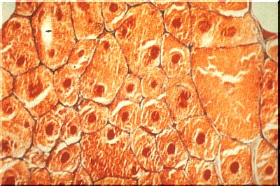 |
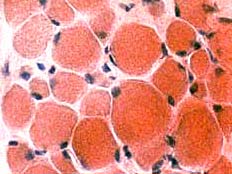
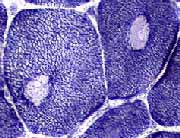
| It derives it's name by the presence of "cores" in the central zones of the myofibers. The cores are best demonstrated by certain special stains (illustrated in trichrome image above), including NADH-TR. In the latter stain, the cores remain unstained. |
 |
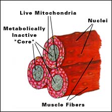 |
| Central Core Disease occurs when the central parts (cores) of some of the muscle cells (fibers) are metabolically inactive, meaning they don't produce enough energy correctly. The cores lack mitrochondria, the energy-producingparts of the muscle cell. |
 |
 |
 |
 |
| Central Core: Pathology |
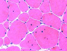
| Myopathic changes in Central Core Disease: Adult (left) & Child (right) * Fiber size: Variability * Connective tissue : Increased * Internal nuclei |
| NADH Stain |
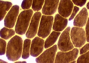
| ATPase, ph 9.4 |
| Central zone, "core", in muscle fibers * Oxidative enzyme activity * Mitochondri Cores run whole length of muscle fiber. |
| Marked type 1 muscle fiber predominencSome cores have loss of central myfibrillar structure. |
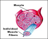
| Cores |
| Muscle fibers |