| CT (3) |
 |
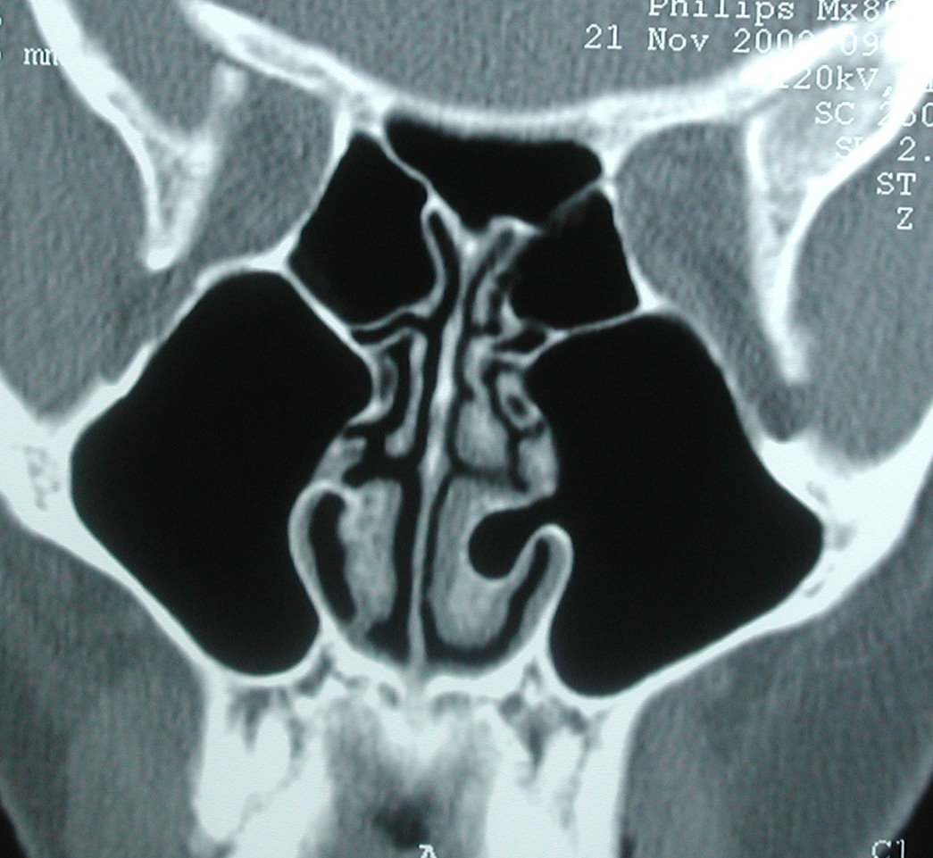
| This patient has maxillary sinus atelectasis. They had sinus disease for many years, and as a result the maxillary sinus shrunk down and "pulled in" the walls of the sinus. The orbit is enlarged as the roof of the sinus came down. Their vision did not suffer though, as the process was slow and symmetric. |
 |
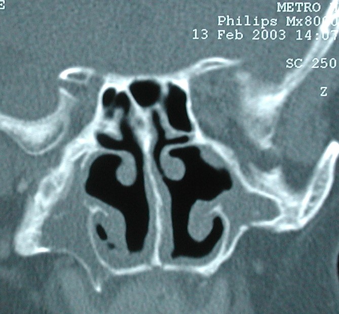
| This CT depicts a fascinating finding: the patient has a concha bullosa of the left inferior turbinate. Furthermore, the IT air pocket connects to her maxiallry sinus. Also (not shown) the nasolacrimal duct drained into the maxillary sinus rather than the nose. They presented with nasal obstruction: a turbinate outfracture was performed AFTER the air cell was opened into the inferior meatus. |