Chinese Medical Journal
1998年4月 第111卷 第4期
| 中华医学杂志英文版 Chinese Medical Journal 1998年4月 第111卷 第4期 |
科技期刊 |
Zheng Xinyu 郑新宇, Guo Kejian 郭克建, Tian Yulin 田雨霖,
Li Jiguang 李继光, Guo Renxuan 郭仁宣 Objective To investigate the cytological pattern and distribution in
nonfunctioning pancreatic endocrine tumors. Chin Med J 1998; 111(4):373-376 It is well-known that pancreatic endocrine tumors (PET) can produce a number of characteristic clinical symptoms due to the hypersecretion of various hormones. Such tumors have been described as ones that secrete insulin, glucagon, gastrin, vasoactive intestinal peptide (VIP), somatostatin, pancreatic polypeptide (PP) and other hormones.1,2 Although many PETs are not associated with clinically significant production of hormones, they are histologically indistinguishable from their functioning counterparts. In general idea, patients are considered to have functional disease if they have compatible clinical symptoms including hypoglycemia caused by an insulinoma, fulminate peptic ulceration by a gastrinoma, diabetes and skin rash by a glucagonoma, etc. The patients are considered to have nonfunctional disease if they do not suffer from hormonally induced symptoms but have an abdominal mass, pain, jaundice, or other abdominal symptoms. Immunohistochemical techniques may sensitively demonstrate hormone production in tumors whether they are associated with hypersecretory syndrome or not. The purpose of this study was to map the cytological pattern of nonfunctioning pancreatic endocrine tumors by immunohistochemistry. METHODS From June 1972 to September 1996, 30 cases of nonfunctioning PET were diagnosed and treated at the First Affiliated Hospital of China Medical University. Surgical resections were performed on all of them. The tumor tissues were fixed in 10% neutral formalin and embedded in paraffin. Routine paraffin sections were stained with hematoxylin-eosin (HE). The immunohistochemical staining for insulin, glucagon, somatostatin, pancreatic polypeptide and gastrin was performed on serial 5-μm paraffin sections using LSAB (labeled streptavidin-biotin) method. The primary antibodies used, their dilution and sources are listed in Table 1. Table 1. Working dilution and source of antibodies |
| Antibody | Dilution | Source |
| Anti-insulin | 1∶200 | DAKO |
| Anti-PP | 1∶700 | DAKO |
| Anti-glucagon | Predilution | DAKO |
| Anti-somatostatin | Predilution | DAKO |
| Anti-gastrin | Predilution | Maxim |
| DAKO: DAKO Corporation (Carpinteria, CA 93013,
USA); Maxim: Fuzhou Maxim Biotech Inc. Normal pancreas (for insulin, glucagon, somatostatin and PP) and gastric antrum (for gastrin) were used as positive control. The following reactions were carried out as negative control: (1) primary antibodies were replaced by phosphate buffered saline (PBS); and (2) primary antibodies were replaced by normal nonimmune rabbit serum as first layer. The staining procedures were as follows. (1) Deparaffinize the sections in xylene; (2) place the sections in absolute ethanol; (3) add 3% hydrogen peroxide,incubate for 5 minutes, and rinse; (4) add blocking serum, incubate for 30 minutes, and tap off excess serum; (5) add primary antibody, incubate for 30 minutes, and rinse; (6) add link antibody, incubate for 30 minutes, and rinse; (7) add diluted streptavidin, incubate for 30 minutes, and rinse; and (8) add substrate solution, incubate for 10 minutes, and rinse.
The cells were classed as positive when they had a rose-red reaction product in the cytoplasm. We counted the positive cells in five high-power (×300) fields and obtained a mean of the results ( RESULTS Clinical features Immunohistochemistry Table 2. Results of immunohistochemical staining |
| Number | % | |
| Total No. of tumors tested | 30 | 100.0 |
| Positive for peptide hormone | ||
| Insulin | 20 | 66.7 |
| Glucagon | 12 | 40.0 |
| Somatostatin | 11 | 36.7 |
| Pancreatic polypeptide | 14 | 46.7 |
| Gastrin | 0 | 0 |
| Negative for peptide hormone | 8 | 26.7 |
| Multihormonal | 17 | 56.7 |
|
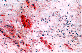
Fig. 1. Immunohistochemical stain for insulin in resected tumor specimen (×300). DISCUSSION We prefer, like Creutzfeldt,1 Heitz et al2 and Mukai et al,3 the term pancreatic endocrine tumors to islet cell tumors. The latter term implies an origin from the islets of Langerhans, which may not always be accurate. Tumors in the pancreas with both exocrine and endocrine components have been reported4 and some authors think that pancreatic duct and endocrine cells originate from the same source. We also avoid another designation "apudoma", because some |
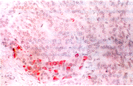
Fig. 2. Immunohistochemical stain for glucagon in resected tumor specimen (×300). |
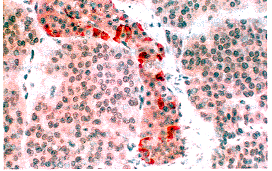
| Fig. 3. Immunohistochemical
stain for pancreatic polypeptide (PP) in resected tumor specimen (×300). Table
3. Positive cells of insulin, glucagon, somatostatin |
| Positive cells | Head | Body-tail |
| Insulin cells | 12.6±5.9* | 40.3±11.8* |
| Glucagon cells | 7.7±5.3* | 31.9± 9.7* |
| PP cells | 33.5±7.4* | 5.7± 2.3* |
| Somatostatin cells | 7.1±1.9** | 9.6± 2.2** |
* P<0.05, ** P>0.05, head group vs body-tail group. |
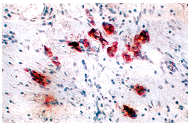
Fig. 4. Immunohistochemical stain for somatostatin in resected tumor specimen (×300). serious questions have been raised about this concept.5 As reported, more than 50% of PETs are multihormonal (56.7% in our study).2,3 Nevertheless, the clinical manifestations are quite different. The most frequent manifestations are always derived from hypersecretion of only one of the hormones produced. A few PETs showed two or more syndromes concurrently or combinations of the above-mentioned hormones,6 and a few other tumors showed transition of one type of symptom to another with the passage of time.7 In some tumors ("nonfunctioning" tumors), hormone production is not clinically evident. The most likely explanation for the variance in clinical manifestations may be the following. (1) Much evidence indicates that the clinical symptoms in multiple hormone-producing PET can often be attributed to the inappropriate secretion of a single hormone. This is confirmed by the finding of positive staining for multiple hormones in which only one serum hormone level was elevated, and the finding supports the notion that the various cell types in PET derive from a single pluripotential stem cell which may differentiate in various directions.3 (2) Although the cells in PET have immunoactivity to multihormones, most or all of the hormones do not have biological activity. (3) Some PETs may secrete a known hormone too small in amount to induce symptoms, produce a hormone with no obvious associated complex of clinical symptoms, secrete a prohormone that is not detectable by conventional methods and is functionally inert, produce a hormone not yet described, or secrete a hormone but fail to release it. The exact mechanism remains to be clarified. Currently it is thought that pancreatic islets arise from immature endocrine precursor cells situated in the epithelium of the developing pancreatic acinar ducts.8 One of the theories of pancreatic endocrine tumor development postulates that PET arise from the totipotential precursor cells.9 The other theory states that these tumors derive from more differentiated islet cells. If the development of pancreatic endocrine tumors was a random event occurring in totipotential cells, it would be expected that the incidence of these tumors would be similar in all portions of the pancreas. Our data, however, showed that positive endocrine cells can be grouped into two distinct anatomic distributions. Most insulin- and glucagon-containing cells were found in the tumors arising from the body and tail of the pancreas; while PP-containing cells usually appeared in the tumors arising from the head of the pancreas. In a recent study with similar result, Sawicki MP et al9 found that approximately 75% of insulinomas and glucagonomas were located to the left of the superior mesenteric artery and no less than 85% of gastrinomas were located to the right of the superior mesenteric artery. Based on the data of Howard et al,10 a bimodal distribution of pancreatic endocrine tumors was identified. In cluster 1 [gastrinomas, pancreatic polypeptide (PP)-secreting tumors, and somatostatinomas], 75% of the tumors were located to the right of the superior mesenteric artery, whereas in cluster 2 (insulinomas and glucagonomas), 75% of the tumors were located to the left of the superior mesenteric artery. The explanation for this regional endocrine cell heterogeneity is the formation of the mammalian pancreas from two distinct primordia, the ventral and dorsal pancreatic buds. Immunohistochemical staining of the mammalian pancreas early in gestation has shown that PP-containing endocrine cells are localized solely in the ventral pancreatic bud, whereas insulin-, glucagon-, and somatostatin-containing endocrine cells are localized in the dorsal pancreatic bud. The small, PP-rich and glucagon-poor islets are the derivatives of the ventral bud primordium, whereas the large, glucagon-rich and PP-poor islets are the derivatives of the dorsal bud primordium. In summary, immunohistochemically, the high positive rate to peptide hormones suggests that the non-functioning pancreatic endocrine tumors are actually not nonfunctioning; they are only asymptomatic pancreatic endocrine tumors. Moreover, an uneven distribution of positive endocrine cells in the nonfunctioning pancreas endocrine tumors within the pancreas was identified. REFERENCES 1. Creutzfeldt W. Endocrine tumors of
pancreas. In: Volk BK, Wellman KF, eds. The diabetic pancreas. New York: Plenum Press,
1977:551. Department of Surgery, First Affiliated Hospital, China Medical University, Shenyang 110001, China (Zheng XY, Guo KJ, Tian YL, Li JG, Guo RX, Zhan Y, Song MM and Shen K) (Received July 24, 1997) 本文编辑: 陶 涛 |