 |
 |
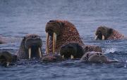
| Introduction Walruses are belong to the class of Mammalian; order Pinnipedia, family Odobenidae, species Odobenus rosmarus. Their males weigh about 800- 1700 kg and females about 400-1250 kg. They are huge animals and adult bulls may approach 2 tons and the famales up to 1 ton. The genus name, Odobenus, "tooth walker", refers to one of their most prominent characteristics, their tusks. These tusks, which are elongated upper canine teeth, are present in both male and females. Tusks are used in mutual display. |
 |
| History & Background A six years old male walrus weighing about 480 kg, living in an Aquatic Animal Park in , Persian Gulf, Iran, was referred to me complaining a draining tract and fistula in the frontal region since 2 years ago. Local vets both in Ukraine and Iran, tried diffrent systemic and local treatment maneuvers. But none of them were successful in subsiding teh problem. Animal was loosing the weigh and appetite progressively. Physical examination adn radiography undr seadtion revieed a draining tract extending from tusk pulp canal following severe abrasion and pulpitis. The problem is fairly known and is common in middle and old aged walruses especially in caged anilams. Following more than a year of search and consult with known aquatic animal's experts, a team was prepared for performing the surgery under general anesthesia. Although the surgical technique was an improtant step, but the most concern of teh team was regarding general anesthesia nad its complications. |
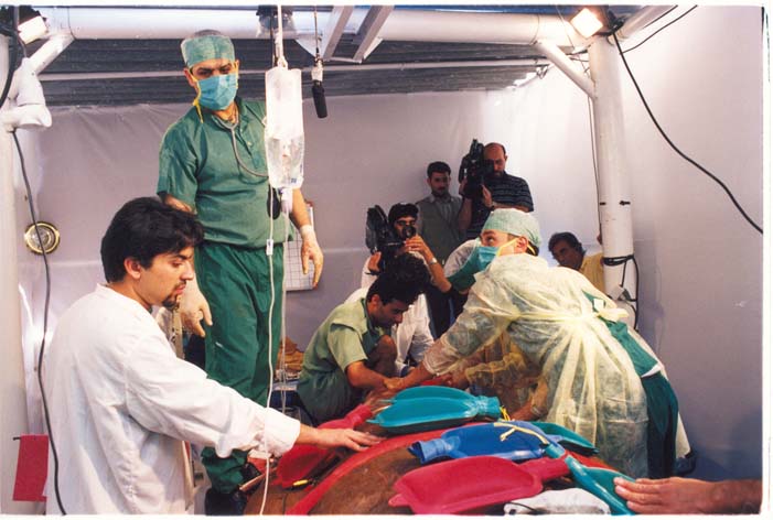
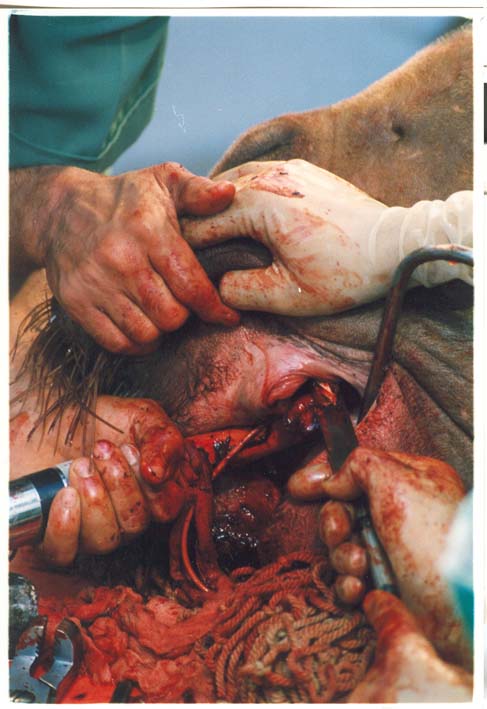
| Technique After preparing most of teh required facilities needed for performing the surgery and anesthesia, the team made the last meetings and the plan was reviewed. A mixture of opioids, centrally acting muscle relaxants and tranquilizers were used for premedication. Dissociatives in combination with thiobarbiturate were administered for induction. After tracheal intubation, anesthesia was maintained by isoflurane. Monitoring during the anesthesia was consisting of ECG monitoring, heart rand respiratory rate recording and measurment of rectal tempearture. In travenous fluids and drugs were administered througgh a veretebral sinus catheter. Surgical technique un brief consisted of cutting the remnant of the tusk, shearing and extraction of thee stump and exploring the hole endoscopically. |
| The picture shows the bloody field of surgery while the endotarcheal tube was kept stable in place. |
| Infected Tusk Extraction in a Walrus First case report from Middle East Region |