Communication in the Body
The Nervous
System Notes
Introduction: Being multi-cellular,
the animal kingdom needed a way for cells to communicate with distant
cells and tissues throughout the body.
WHY?
SURVIVAL! All of the different specialized
cells in the body must be managed and coordinated to maintain homeostasis
and to sense and react to a threatening environment.
Communication between tissues took two forms:
1. The Endocrine
System uses chemical signals (like sending a letter by mail)
2. The Nervous System uses electrical signals
(like making a phone call).
I.
The Endocrine System:
Parts of the endocrine
system:
A. Endocrine Glands: This type of gland produces chemical messengers called hormones,
releasing them directly into the blood stream.
B.
Importance of the hormones: The hormones
(chemical messengers) attach to target cells signaling them to function
in a certain way.
1. Hormones maintain homeostasis in the
body by controlling metabolism; body temperature; growth and
development; reproductive systems; and levels of salt, water, calcium, and
glucose in the blood.
2. Hormones = slow communication: Being
released into the blood, hormones must travel throughout the body before
reaching its target cells.
3. Hormones = widespread communication: Dumping hormones
in the blood is like sending out many letters or flyers in the mail. Although slow, the letters (hormones)
communicate with many tissues at the same time.
C.
The Pituitary gland: Located at the base of the brain,
the pituitary gland releasing hormones that controls all other glands.
The pituitary gland is connected to the part of the brain called the hypothalamus.
1. Function of the Hypothalamus: By monitoring
the blood flowing through the brain the hypothalamus knows when to
signal the pituitary gland to release hormones to maintain
homeostasis in the body.
II. The Nervous System:
Parts of the Nervous System:
A.
The Nerve- a bundle of nerve cells.
The Neuron is the basic unit of
function of the nervous system. It
is a specialized cell that carries electrical messages
called impulses throughout the body.
1. The
Structure of a Neuron: (A Motor Neuron)
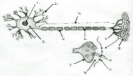
- Nucleus- control center of the cell
- Dendrites-
extensions that bring impulses to the cell body. There are many of them to receive
impulses from all directions.
- Cell Body- processes incoming impulses and sends them out.
- Axon- usually one large extension that carries impulses away from
the cell body to another nerve cell or tissue.
- Myelin sheath- lipid
insulator found on some axons.
It helps transmit impulses faster.
- Node- spaces between the myelin where the axon is exposed. Impulses jump from node to node.
- Axon ends- places where electrical impulses from one neuron must be
changed into chemical signals to be transferred to another neuron
or tissue.
- Muscle- In the case of a motor neuron, impulses can stimulate
muscle tissue to contract.
- Synapse- gap or empty space between two neurons or between a neuron
and tissue. Two neurons do not
actually touch each other.
- Neurotransmitters- chemical signalers that allow impulses
to jump the synapse to a new neuron or tissue.
- Receptor (gated ion channel) – place where neurotransmitters bind to
the new neuron or tissue.
2. Three
types of neurons (nerve cells):
a.
Sensory neuron – carry
impulses from the sense organs/cells to the spinal cord and brain.
b.
Motor neurons – carry
impulses from the brain and spinal cord to the muscles or organs.
c.
Interneurons – processes
impulses. Interneurons are found
in the brain and spinal cord connecting sensory and motor neurons together.
2.
Motor and sensory neurons can reach as long as a meter
in length.
3.
The nerve:
Neurons do not work alone! A bundle
of neurons surrounded by connective tissue and blood vessels
make up a nerve.
B.
The Central Nervous System (CNS)
1. The two main parts of the Central nervous system
are the Brain and
Spinal cord.
2.
Three membranes called the meninges
protect the brain and spinal cord. A
fluid called the cerebrospinal fluid fills the gap between the
membranes to cushion against injury.
3. Structures of the central nervous system:
1.
Brain- control
center for the entire nervous system
2.
Spinal cord- primary
link between brain and body. It
processes some impulses.
3.
Cerebrum- largest
part of the brain responsible for intelligence, personality, learning and
judgment.
4.
Cerebellum- second largest part of the brain
that controls balance and coordination.
5.
Medulla Oblongata
(brainstem)- controls involuntary actions like blood pressure, heart rate,
breathing, and swallowing.
6. Spinal Nerves- connects the spinal cord to the rest of the body.
C.
The Peripheral Nervous System (PNS)
1. Includes all nerves and associated cells that connect the brain and spinal cord to the rest of the body.
2.
The Two Divisions of
the PNS:
a.
Sensory Division- receives
messages from the environment, sending them to the spinal cord and
brain.
b.
Motor Division- sends
impulses from the brain and spinal cord to the body. Two subdivisions:
1.
Somatic= includes
the nerves that control voluntary skeletal muscle movements.
2.
Autonomic= includes
the nerves that control involuntary actions of organs like the beating
of the heart and contracting of stomach muscles.
3. A reflex is an automatic (unthinking) response to a stimulus. These are controlled by the spinal cord instead of the brain.
4. The Reflex Arc Example:
1.
heat receptor
2.
sensory neuron
3.
interneuron
4.
spinal cord
5.
motor neuron
6.
muscle
D. The Senses
1. The largest sense organ in the body is the skin.
a.
The skin has receptors scattered throughout the skin.
b.
These receptors include: heat receptors, separate cold
receptors, light touch receptors, pressure receptors, and pain
receptors (free nerve endings).
2.
Taste and Smell
are chemical senses:
a.
The tongue has sensory receptors called taste buds.
They can detect four major tastes: sour, salty, bitter, and sweet.
b.
Smells are
picked up using sensory receptors on the roof of the nasal cavity. These messages travel directly to the brain
by way of the olfactory nerve.
3. Hearing and balance:
a.
The ear has three main parts: the outer, middle, and
inner ear.
b.
**The auditory canal ends with the tympanic membrane or
eardrum, which vibrates, passing on the vibrations to three tiny bones in
the middle ear called the hammer, anvil, and stirrup. Vibrations then travel trough the
fluid-filled cochlea of the inner ear.
There, hair-like sensory receptors send impulses along the
auditory nerve to the brain where we recognize it as sound.
c.
Balance: Three tiny fluid- filled
semicircular canals sense changes in the position of the head,
helping to maintain balance.
d. Parts of the Ear:
1.
auditory canal
2.
eardrum- tympanic membrane
3.
hammer-bone
4.
anvil-bone
5.
stirrup-bone
6.
cochlea
7.
auditory nerve
8.
semicircular canals (3)
4. Vision- the eye
a. The transparent convex shaped lens focuses light
on the back inner part of the eye. The
light hits a layer of cells that are very sensitive to light and color called
the retina. This part has
receptors called cones, which are sensitive only to bright light,
and help distinguish form and color.
The other structures called the rods are sensitive in dim
light enabling people to see shades of gray.
b. Other parts of the eye: cornea, the clear outer
covering; the iris, the colored part; the pupil,
the opening into the inner eye; the humor, the fluid part of the
eye; and the sclera, the white of the eye.
c. The optic nerve carries impulses to the
brain where the images that were inverted by the convex lens are interpreted as
being right side up again.
d.
Parts of the Human Eye:
1.
cornea
2.
iris
3.
pupil
4.
lens
5.
humor
6.
optic nerve
7.
retina
8.
sclera