(S. A. Patney: Strabismology Desk Reference, chapter 47, JKA Publications)
DOUBLE ELEVATOR PALSY (Monocular Elevation
Deficiency)
As there are varied causes of absence of elevation of
an eye, including nonparalytic ones, the term "Monocular elevation deficiency"
(MED) is more apt. Apparently in such cases it appears as if both the elevators (the
superior rectus and the inferior oblique) are not functioning but recently it has been
shown that a paralysis of superior rectus muscle alone can cause a deficiency of elevation
in abduction as well as in adduction. This is due to the fact that superior rectus is the
main elevator of the eye and elevates the eye not only in abduction and primary position
but also in adduction.1, 2 The inferior oblique is the main extortor. However,
there are some cases in which there is true palsy of both the elevators resulting from a
supranuclear involvement affecting upgaze.
Definition
Double elevator palsy or monocular elevation deficiency refers to a
condition in which the eye can not be elevated in abduction, adduction or from primary
position. It is associated with ptosis.
History
1864: Bilateral upgaze palsy described first by Henoch3
1883: Perinaud described upgaze, downgaze and total vertical gaze
palsies3.
1942: White4 reported congenital paralysis of elevation
associated with hypotropia and ptosis of the affected eye. The deficiency of elevation was
thought to be due to paralysis of both, superior rectus (SR) and inferior oblique (IO).
1954 Dunlap5 named the condition as "double elevator
palsy".
Prevalence
Double elevator palsy is not a common problem but it is certainly
seen much more frequently than double depressor palsy or DDP (also termed as monocular
depression deficiency or MDD). Exact figures are not available.
Terminology
Double elevator palsy is also known as "Monocular elevation
paresis" or "Monocular elevation deficiency".
Classification
The following is a modified classification of Double elevator
palsy (DEP) or monocular elevation deficiency (MED):
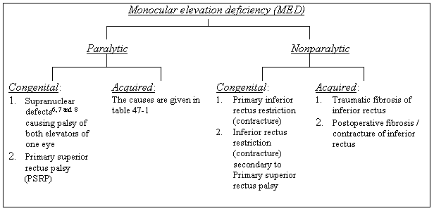
NOTE: Congenital cases occur sporadically. Acquired cases have a
varied etiology. Thorough systemic and neurological examination is necessary to find the
cause.
Etiology
In these cases it is presumed that there is an interruption of
supranuclear input from riMLF10 (rostral interstitial nucleus of Medial
Longitudinal Fasciculus that mediates upgaze) into III CN nucleus. Various causes3
of congenital and acquired DEP have been mentioned in the table 47-1 on page 907.
It has been shown relatively recently1 that superior
rectus is the main muscle for elevation of the eye, be it in abduction, adduction or
primary position from where the eyes are elevated. In some of the cases the defective
elevation can therefore be explained by the presence of a marked superior rectus palsy
alone.
To understand the etiology a basic knowledge of anatomy of
supranuclear pathways is necessary. The main points are given in the following text:
- The supranuclear pathways for upgaze are situated in the pretectum in
midbrain.
- The important structures are rostral interstitial nucleus of the
medial longitudinal fasciculus (riMLF) and the posterior commissure.
- The efferent fibres for upgaze leave the riMLF and decussate in the
midline of the posterior commissure, pass through the pretectum and enter the subnucleus
of the superior rectus (SR) in the oculomotor nucleus.
- After leaving the SR subnucleus the upgaze fibres to the SR cross the
midline again.
- After the double decussation of the upgaze fibres the SR is
innervated from the ipsilateral riMLF and from the contralateral pretectum and the SR
subnucleus.
- According to a hypothesis the supranuclear fibres for up and down
gaze may be compartmentalized9. This borne out by the fact that one may come
across cases in which saccadic recordings may be found to be abnormal in upgaze and normal
in down gaze.
Table 47-1:
| Type of DEP or MED |
The etiological factors |
|
Congenital
|
1
Supranuclear defects6, 7 and 8 causing palsy of both elevators of one eye |
| 2
Primary superior rectus palsy (PSRP) |
| 3 Primary
inferior rectus restriction (contracture) |
| 4
Inferior rectus restriction (contracture) secondary to Primary superior rectus palsy |
| 5.
Neonatal hypoxia is held responsible in some cases.11 |
| Acquired |
1
Cerebrovascular disease (hypertension, arteritis, thrombo-embolism) leading to acute
diplopia, unconsciousness, neurological symptoms |
2
Midbrain neoplasms (Acute diplopia):
Peneocytoma
Acoustic neuroma
Metastatic tumors |
| 3 Sarcoidosis |
4 Infective
diseases |
| 5 Venereal
disease (Syphilis) |
6 Other causes |
Symptomatology / Clinical Picture
The typical clinical picture of MED has the following components:
- Associated systemic symptoms
may be present in acquired MED
according to the type of involvement.
- Ocular symptoms
: Typically, no symptoms in congenital MED. In
acquired MED however, vertical diplopia is present in elevation (upgaze) only, none in PP.
Diplopia in PP is there only if the affected eye is hypotropic in PP also. In acquired MED
the onset of diplopia is acute and it is present in PP and elevation.
- Compensatory Head Posture (CHP)
: Various types of CHPs have been
seen with MED (DEP). If binocular vision is present a chin elevation by tilting the head
back is usually seen. Head posture may be normal if there is no hypotropia in PP or if the
hypotropic or the hypertropic eye is amblyopic.
Ptosis: A hypotropic eye shows ptosis because of the fascial
attachments between the levator palpebrae superioris and the superior rectus muscle.
Usually it is a pseudo-ptosis that disappears when the hypotropic eye is made to take up
fixation in primary position. Ptosis is most noticeable when the non-affected eye fixes.
As the hypotropic eye moves up to primary position to fixate, the ptosis disappears if
there is no true ptosis.
Presence of true ptosis must be ruled out by covering the normal
eye. It may be present in about 50% cases12. In these cases when the patient is
made to fix with the affected eye the ptosis recovers only partly.
Marcus Gunn phenomena may be present12 with the ptosis.
DEP may also be associated with other complications in some patients with intracranial
involvement.
- Deviation: Hypotropia
of the affected eye when normal eye
is fixing; hypertropia of the normal eye when the affected eye is fixing.
Usually it is the hypotropia of the affected eye and the normal eye fixates. But if the
normal eye is amblyopic (and sometimes if the hypotropic eye is the dominant eye and is
preferred for fixation) it may be the constantly deviating eye with hypertropia.
- Monocular limitation of upgaze
above midline (horizontal plane)
is present in all the three horizontal positions, i.e., abduction, midline position and
adduction.
- Bell'
s phenomenon is usually present unless inferior rectus has
developed contracture.
- Amblyopia:
Amblyopia may be present if there is a constant
deviation or if there is anisometropia. A hypotropic eye is usually not found to be
amblyopic because depression is the most often used position. Presence or absence of
amblyopia depends on fixation preference. If the non-paretic eye is the constantly
deviating (hypertropic eye it may develop amblyopia. If there is no fusion present the
hypotropic paretic eye is likely to become amblyopic.
Characteristic signs of supranuclear MED:
- It is generally congenital.
- Vertical movements above the horizontal plane are absent in the
affected eye in abduction, midline and adduction. It is a monocular deficiency of
elevation.
- Forced duction test is negative, meaning thereby that there is no
resistance to forced (passive) elevation13 of the affected eye.
- Bell's phenomenon is present indicating a normal III cranial nerve
(oculomotor) nucleus , fasciculus.6, 13
- Saccadic velocity in vertical direction is normal or only slightly
reduced below the horizontal plane but absent above it (in upgaze).
Examination
The following points are particularly important in the examination
of cases of MED:
- History
: It is important to take a thorough history in order to
find out whether the MED is congenital or acquired. If acquired one has to decide the
cause by taking history of systemic diseases, malignancy etc.
- Pupillary examination
should be carried out to rule out /
identify anomalies of pupil (important in neurological disorders).
- Cover test
: It should be done in all the three vertical planes,
fixing each eye. In elevation there is marked hypotropia of the affected eye. In
PP no deviation to some hypotropia and in depression no vertical deviation. There may
be a co-existent horizontal deviation.
- Ocular motility examination
: Both versions and ductions should be
tested in the 9 cardinal directions of gaze. A special feature of MED is an equal degree
of defect of elevation in adduction as well as in abduction.
- The Park's three-step
test and Bielschowsky's head tilting
test is carried out to distinguish MED from superior oblique palsy.
- Bell'
s phenomenon, if present means the palsy is supranuclear.
- Examination on a major amblyoscope
is useful in estimating the
grade of binocular vision (SMP, Fusion and its range and stereopsis) present, measuring
the deviation (vertical, horizontal and torsional) in various directions of gaze and
determining the presence, type, direction and degree of suppression. It is usual to find
suppression in upgaze and sometimes in PP also. Good binocular functions are generally
present in downgaze with no suppression. Angle of deviation fixing each eye in turn,
should be measured in the 9 cardinal directions of gaze.
- Stereo-tests
: Results on Titmus / Wirt fly test, TNO test etc
give a fair idea of the state of binocular vision even if major amblyoscope is not
available in a clinic.
- Worth Four Dots test
also helps in estimating the state of
binocularity, presence / absence of suppression, presence, directions and type of
diplopia.
- Prism Bar cover test
will give a measurement of the deviation in
various cardinal directions of gaze, particularly in upgaze, PP and downgaze.
- Forced duction test
(FDT): In case of primary palsy the FDT is
negative while in cases of MED due to contracture of inferior rectus, it is positive.
- Active force generation
test is done to decide the severity of
palsy. One should be aware of the possibility of contracture of inferior rectus (IR).
- If recording of saccades is not possible, they can be tested
clinically without any equipment. The patient is asked to look to and fro between the
PP and the upgaze (to test saccades in upgaze) and to and fro between PP and downgaze to
test saccades in downgaze. If there is a primary paresis there are floating saccades. In
the cases of restrictive strabismus and in supranuclear MED the saccades are rapid and
they stop suddenly.
- Objective recording of saccades
is done by electro-oculography or
scleral search coil technique, the latter being more accurate. Vertical saccadic
velocity is of help in differentiating between MED, inferior rectus (IR) restriction and
SR paresis.
- Hess chart
gives an idea of the amount of deviation in primary
position and the degree of underaction in various fields, e.g., elevation in adduction and
elevation in abduction.
- Field
of BSV: It helps in deciding the extent BSV.
- Radiological
diagnosis: CT scan and MRI of orbits help in
diagnosing the missing and defective muscles (e.g., contracture). In cases of acquired
palsy with systemic, particularly neurological complications, the patient must be referred
for a thorough neurological examination.
Diagnosis
NOTE: The diagnosis of congenital MED is made clinically. No
laboratory and radiological tests are necessary. However, they can be performed for
academic and research purposes.
Acquired MED needs further tests to determine the cause.
The following points are particularly to be kept in mind while
making a diagnosis of MED:
- If the patient is a child the parents will usually be able to tell
the age of onset.
- Presence of amblyopia indicates a long duration and therefore point
to the presence of a congenital MED.
- In most cases the supranuclear MED
is congenital. Bell's
phenomenon is present indicating a normal III CN, fasciculus and nucleus14.
- Presence of systemic disorders
like cardiovascular disease,
malignancy etc will be in favor of acquired palsy.
- Presence of neurological signs
like loss of consciousness,
vertigo, ataxia, tinnitus, deafness and others will go in favor of acquired palsy of SR. A
thorough neurological examination is a must in these cases.
- Positive forced duction test
on the direct antagonist of superior
rectus (SR), that is, the ipsilateral inferior rectus (IR), will indicate the presence of
contractures / tightness of IR leading to secondary SR restriction. If the inferior rectus
is tight and fibrotic SR will not be able to move the eye up satisfactorily.
- If there is no primary palsy of SR the saccades will be normal
otherwise the saccadic velocity will show an upward slowing in upgaze, may be in PP
and will be normal / or show mild slowing in down gaze.
- Radiological tests like plane (x-rays) radiogram, CT scan and MRI of
skull / orbits
may be of great help, even in cases of congenital MED if the cause is a
missing SR or the IR is fibrotic.
- Neurological (Neuro-ophthalmological) examination is a must in all
cases of acquired MED.
Table - 2, Differential diagnosis of congenital Vs acquired MED
Congenital
(supranuclear) MED |
Acquired
MED (DEP) |
| 1. Present since birth |
1. Can start at any age depending
upon the cause but more often in older age group with diabetes, hypertension,
atherosclerosis, arteritis, other CV disorders |
| 2. No complaint except ptosis /
strabismus |
2. Diplopia is a common
complaint. Ataxia, tinnitus, loss of consciousness, palsy of other oculomotor nerves may
be present |
| 3. No relevant systemic disease
is present |
2. Systemic diseases as above
present, specially in older patients |
| 4. Neurological examination is
negative |
3. Positive more often than not |
| 5. Patient is usually a child |
4. Patient is usually an adult,
more often in older age group |
| 6. Amblyopia and suppression
common |
5. Amblyopia and suppression
rare. Diplopia common |
Legend: See below
Table 3, Differential diagnosis of congenital MED
Name of
the disorder |
Distinguishing
features |
| 1. Third CN palsy (Figure 1) |
True ptosis, XT, limitation of
elevation, depression and adduction, pupillary signs may be present. |
| 2. Vertical retraction syndrome
(like Duane's retraction syndrome) |
Eye retraction in downgaze and
strongly positive forced duction test indicating fibrosis of IR |
| 3. Congenital aplasia of
superior rectus w or w/o congenital aplasia of inferior rectus |
Association with
craniofacial dysostoses/anomalies common. In IR aplasia
absence of depression. |
| 4. Anomalous insertion of
inferior rectus w or w/o anomalous insertion of superior rectus |
No definite distinguishing
features. The clinical picture varies from case to case. |
| 5. Congenital fibrosis of
inferior rectus |
Strongly positive forced duction
test: eye can not be elevated even passively, indicating fibrosis of IR. |
Legend: CV = Cardiovascular; IR = Inferior rectus; XT = Exotropia; w = With;
w/o = Without; CN = Cranial nerve
Table 4, Differential diagnosis of acquired MED
Name of
the disorder |
Diagnostic
features |
| III CN palsy, superior
division |
Ptosis, vertical diplopia,
hypotropia. Sparing of pupil and EO muscles supplied by inferior division of III CN, IV
and VI CN. Forced duction test (FDT) negative. Force generation test: weak or no force
generation (FG). |
| Post-cataract surgery SR palsy |
SR paresis due to myotoxic
effects of local anesthesia. Forced duction test excludes fibrosis of ipsilateral
inferior rectus. SR tightness may be revealed. History of cataract surgery + relation with
symptoms. FG: weakened or normal. |
| Orbital floor fracture with IR
entrapment |
History of injury, diplopia,
enophthalmos, anesthesia of V CN, second division, FDT +, FG: OK, fracture / entrapped
inferior rectus (IR) seen on CT scan / MRI |
| Orbital cellulitis |
History of sinusitis +/-,
pain in orbital region, inflammation / swelling of lids, exophthalmos, systemic symptoms
like fever, decreased vision, Limitation of ocular motility
not limited to elevation. CT scan abnormal. |
| Orbital tumors, upper part |
Proptosis with globe pushed down,
limitation of other movements also may be present |
| Progressive external
ophthalmoplegia |
Bilateral, progressive defect of
ocular motility with variable degree of involvement of various muscles, ptosis, inferior
and medial recti affected more often and in earlier stages, FDT negative, FG
weakened/normal, decreased saccades, abnormal EMG, ragged red fibres on biopsy. |
| Myasthenia Gravis |
Ptosis (worse after fatigue),
diplopia, progressive defect of ocular motility with variable degree of involvement of
various muscles, orbicularis weakness, FG weakened, FDT negative. Tensilon test positive.
Abnormal EMG. |
| Thyroid orbito-oculopathy |
History of thyroid disease,
exophthalmos, retraction of upper lid, lid lag, congestion with dilatation of blood
vessels on EOMs, restricted ocular motility with variable degree of involvement of
different muscles, FDT positive specially during stage of fibrosis of muscles, swollen
muscles seen on CT scan/MRI |
| Cerebellar tumors |
Ocular flutter, papillaedema,
progressive ataxia, abnormal OKN, neurological exam. / CT scan of brain / brain-stem may
show SOL. |
| Labyrinthine disorders |
Acute onset of vertigo,
nystagmus, tinnitus, nausea, abnormal OKN, neurological work-up and CT scan / MRI of brain
may be positive. |
Legend: FDT = Forced duction test; FG = Force generation; EMG =
Electromyogram; OKN = Optokinetic nystagmus; SOL = Space occupying lesion; SR = Superior
rectus; IR = Inferior rectus; CN = Cranial nerve.
Figure 1, III CN Palsy OS:
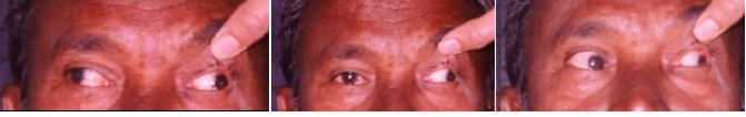
Legend: DV = dextroversion; LV = levoversion; ++ = overaction;
LXT = Left exotropia; PP = Primary position.
Treatment
- Correction of refractive error
- Treatment of Congenital MED
- Treatment of acquired MED
Treatment of Congenital MED
Treatment of Amblyopia
Treatment of significant CHP
Treatment of deviation (hypotropia)
Treatment of ptosis
- Treatment of Amblyopia
: The amblyopia can be of various types,
namely strabismic, anisometropic and ametropic (binocular). The treatment is carried out
in conventional way. The main points are as follows:
A. Strabismic and anisometropic Amblyopia
- Upto the age of 7-8 years
every case of strabismic /
anisometropic amblyopia
should be treated by conventional occlusion provided the fixation
is central and refractive error has been corrected.
- Above the age of 8 years (visual maturity):
- Strabismic amblyopia with central fixation
is treated only if
binocular functions (fusion with a definite range) are present.
- Anisometropic amblyopia with central fixation
and fusion is
treated by occlusion as usual. I have found CAM useful in quite a few of these cases.
- Amblyopia with eccentric fixation
is treated conventionally only
if the patient is a young child (upto the age of 2-3 years). These cases are also divided
into 2 types:
- If the eccentric fixation is fixed
(well established) at one
point inverse occlusion is given for 6 week and then surgery is performed. If
however, the fixation changes after the 6 weeks, conventional occlusion can be
tried to see if the fixation improves (becomes unsteady or moves nearer to macula).
- If the eccentric fixation is
unsteady, conventional occlusion is
given a chance to see if it improves. If it does not, inverse occlusion for 6 weeks is
followed by surgery.
B. Treatment of ametropic amblyopia
- Adequate correction of refractive error and constant use of glasses
can gradually lead to improvement of visual acuity in young children.
- Use of yellow glasses has been credited with improvement of visual
acuity in cases of ametropic amblyopia without any strabismus of anisometropia. We are
carrying out a study on use of yellow glasses in ametropic amblyopia and await the
results.
- Treatment of significant CHP
: As the CHP is adopted because of a
hypotropia in PP, surgery is indicated to correct it. It should be carried out
while the patient is still young so that any deformities in the spine that have been
brought about by a long-standing CHP can be corrected. Significant degree of elevation of
chin can lead to antero-posterior changes in spinal curvature (while a head tilt can cause
a sideways change).
- Treatment of deviation (hypotropia)
: Surgery has to be
resorted to. It is only indicated if it is present in PP and can be a cause of amblyopia,
loss of fusion, pseudoptosis and CHP (and possibly spinal changes). If there is no
hypotropia in horizontal plane and in downgaze, no active treatment is required. The
patient is called for checks from time to time to see if the deviation is becoming
manifest.
- Treatment of ptosis
: It is only needed if there is true ptosis
present that poses a cosmetic problem. A pseudoptosis is automatically corrected when
hypotropia is taken care of. There may be both factors present and if ptosis is not fully
corrected and cosmetically acceptable after the hypotropia is gone or significantly
reduced (to become a hypophoria), ptosis correction is carried out by surgery.
Treatment of acquired MED
- Conservative
- Medical and neurological
- Orthoptic for suppression and amblyopia
- Observation for any signs of change for better or worse
- Surgical
As there is a wide variety of causes of acquired MED, thorough
investigations (general, ophthalmologic, neurologic (neuro-ophthalmologic and orthoptic
(ocular motility work out) have to be undertaken. In a case of recent palsy one has to
wait for at least 6 months to allow for spontaneous recovery.
The main points are given below:
- The underlying systemic neurologic conditions must be treated first.
- Observation is the best policy. Surgery should not be carried out as
long as there is spontaneous recovery. A period of six months after the onset of palsy
should elapse before surgery is resorted to.
- The indications of surgery
in acquired MED are as follows:
- Vertical deviation in PP
- Persistent vertical diplopia in PP
- Suppression
- Amblyopia
- Small field of BSV
- Ptosis correction
by surgery should only be done if it persists
after the vertical deviation (and hence the pseudo-ptosis) is eliminated.
The aims of surgery
- To increase the field of BSV by achieving orthotropia with fusion in
as large a field as possible. The field of BSV should preferably be centered in primary
gaze (PP).
- To eliminate any significant degree of CHP (chin elevation) by
achieving BSV in PP.
- To eliminate hypotropia in PP (and depression where diplopia is
hardly ever present)
- To treat ptosis, which is usually pseudoptosis due the presence of
hypotropia in PP. Eliminating hypotropia gets rid of it.
- To eliminate manifest deviation (hypotropia with or without
horizontal deviation)
Pre-requisites of surgery for acquired MED
- There should be no change in the condition of MED for a few weeks on
two consecutive checks.
- At least 6 months must have elapsed after the onset even if the
condition is stable.
- Systemic conditions like diabetes and hypertension must be under
control.
- Neurologic conditions like encephalitis, meningitis, and intracranial
space occupying lesion (SOL) etc. should have been taken care of.
- Condition of stroke patients should have been stabilized.
- Suppression and amblyopia should have been attended to. They occur
only after a significant length of time, specially the latter.
Precautions before surgery
- The patient / parents must be explained, particularly in cases of SR
palsy and supranuclear palsy that full elevation may not be restored. In some cases no
improvement in elevation (upgaze) is possible.
- The patient / parents must be informed that more than one operations
may be required for the strabismus.
- The patient / parents must be cautioned that ptosis correction by
surgery may be required later if eliminating hypotropia fails to get rid of it.
Techniques of surgery
- If the forced duction test is positive
for restriction of the
ipsilateral inferior rectus (IR) due to fibrosis / contracture: These cases fall into two
groups, namely 1. Those with primary IR restriction / tightness / contracture / fibrosis
and 2. Those with IR restriction secondary to SR palsy.
Group 1 cases: recession of IR (5-8 mm) with recession of
conjunctiva (4 mm) is indicated. Good results have been reported14 after this
procedure.
The surgery on a tight rectus muscle is difficult and risky. Three
serious complications that should be guarded against are perforation of the globe,
avulsion of inferior rectus if it is pulled with force and slippage of the muscle
(particularly if pre-placed sutures are not applied).
Another precaution during surgery on IR is that connections
between the IR and the lower lid should be dissected thoroughly to avoid a postoperative
lower lid retraction.
Forced duction test is repeated after the IR is disinserted
(detached) from the globe. The eye should now elevate freely if all the adhesions and
tight IR fibres have been severed.
Finally the forced duction test is repeated after the IR is
reattached. Elevation of the eye by at least 20-25 degrees14 should be
possible.
Group 2 cases: Fibrosis of the IR in these cases is secondary
to SR palsy. Therefore IR recession alone may often be inadequate to eliminate hypotropia
in PP and Knapp's procedure15, 16 has to be performed in addition to the IR
recession. It is safer to perform Knapp's procedure in the second stage after 4-5 months
to reduce the risk of anterior segment ischaemia. An alternative is to do them in one
sitting with the sparing of the ciliary vessels to the recti. It is easier in case of
vertical rectus muscles than in the case of horizontal rectus muscles.
.
NOTE: In Knapp's procedure15, 16 insertions of
ipsilateral MR and LR are vertically transposed to that of SR.
- If the forced duction test is negative
for restriction of the
ipsilateral inferior rectus (IR): In these patients there is either a SR palsy or
supranuclear MED. In both these conditions Knapp's procedure15, 16 is
indicated. This technique causes insignificant improvement in upgaze but corrects about 20
to 35 PD of hypotropia6, 15, 16, 17, 18, 19, 20 in PP. For various degrees of
hypotropia the effect of the Knapp's procedure can be graded by using it after IR
recession and / or using the various modifications of the Knapp's procedure as suggested
below:
- Hypotropia of 10 PD or less in PP
: There are two options
depending upon the degree of elevation above midline: A) If there is no elevation
possible above midline: partial tendon transposition of horizontal rectus muscles
(modified Knapp's procedure) without any previous surgery (e.g., IR recession).
B) If there is some elevation present above midline:
resection of SR21 (as an alternative to horizontal rectus muscles
transposition) with or without one of the other secondary procedures may deliver the
desired result.
- Hypotropia of 25 PD or less
: IR recession followed by partial
tendon Knapp's procedure can take care of the hypotropia.
- Hypotropia of 25-35 PD
: IR recession followed by full tendon
vertical transposition of horizontal rectus muscles near the SR tendon is recommended. As
already mentioned, the two procedures should be performed in two sittings to avoid
anterior segment ischaemia. However, if the two procedures are to be done at one session
sparing of ciliary vessels to the recti is done.
- Hypotropia of more than 35 PD in cases of MED without IR restriction:
Classic Knapp's operation with vertical transposition of the full horizontal rectus
muscles to the SR insertion + posterior fixation suture on the transposed muscles21
has been advised.
Ptosis correction
If there is true ptosis present it will persist after the correction
of hypotropia. These are the cases that need surgery for ptosis correction. A resection of
the levator muscle by external route is indicated if there is some function in that
muscle. One must also make sure that there is some function in the SR and the eye can be
raised at least partially, to avoid the risk of exposure keratitis.
Complications and problems after surgery for MED
Names of the main problems that can occur in the postoperative
period are given below:
- Undercorrection
- Overcorrection
- Worsening of diplopia
- Anterior segment ischaemia
- Horizontal tropia
- Retraction of lower lid
Undercorrection: If there is residual hypotropia after the
Knapp's procedure one should wait and watch, as improvement has been reported15
after some time. The management strategy may be devised21 as follows:
- After Knapp's procedure: Recession of ipsilateral IR
can be done,
particularly if there is even the slightest indication of tightness in the muscle.
- An alternative to recession of the IR, is a recession of the SR of
the other eye
. A plus point of this procedure is that it lessens the incomitance in
the upgaze that s due to underaction of SR / MED.
- If Knapp's procedure has not been performed and only a recession of
IR has been done in the first stage, a partial horizontal rectus muscles vertical
transposition
is indicated.
Overcorrection: In case of overcorrection leading to hypertropia
and advent / worsening of diplopia, the problem is not likely to improve. According to
reports the hypertropia tends to get worse15, 17 with time. These cases are
managed as follows:
- If the overcorrection is a result of Knapp's procedure
: The
insertions of the lateral and medial rectus muscles that were supraposed (transposed
superiorly) are lowered.
- If the IR recession had been performed
: the IR id advanced by 1mm
for a hypertropia of 3 PD, 2mm for 6 PD of hypertropia and so on.
Worsening of diplopia: This situation develops if the IR on the
affected side has been recessed excessively leading to a weakness of the eye in downgaze
as compared to the other eye. Thus a hypertropia of the operated eye is created in
downgaze, the IR on the sound side lowering the eye more than the operated IR on the
operated side. The usual procedure advised for these patients is recession of
the IR of the sound unoperated eye to match the position of the two eyes in downgaze.
If however this procedure does not produce the desired amount of weakening and the
diplopia persists in downgaze, prismatic correction is prescribed.
Anterior segment ischaemia: It may result if more that two
rectus muscles are operated upon at one sitting. To prevent this complication a ciliary
vessels sparing procedure may be carried out.
Horizontal tropia: It may be produced if the two horizontal
rectus muscles were not transposed equally.
Retraction of lower lid may result if the attachments between
the eyelid and the IR that was recessed were not severed sufficiently.
References
- Boeder, P.: The co-operation of extraocular muscles, Am. J.
Ophthalmol. 51:469, 1961.
- Jampel, R.S.: Extraocular muscle action from brainstem stimulation of
Macaque, Invest. Ophthalmol. 1:565, 1962.
- Ziffer, A.: Clinical strabismus management: Principles and surgical
techniques. Editors: Rosenbaum, Arthur L. and Santiago, A.P., 1999, Philadelphia, PA, W.B.
Saunders Company, p. 272.
- White, J.W.: Paralysis of the superior rectus and the inferior
oblique muscles of the same eye, Arch. Ophthalmol. 27:366, 1942.
- Dunlap, E.A.: Diagnosis and surgery of double elevator palsy, Trans.
Am. Ophthalmol. Soc. 3:1554, 1952.
- Barsoum Homsy, M.: Congenital double elevator palsy, J. Pediatr.
Ophthalmol. Strabismus 20:185, 1983.
- Bell, J.A. et al: Congenital double elevator palsy in identical
twins, J. Clin. Neuroophthalmol. 10:32, 1990.
- Hitz, J.B.: Discussion: Paralysis of the superior rectus and the
inferior oblique muscles of the same eye, 27:366, 1942.
- Kirkham, T.H. and Kline L.B.: Monocular elevation paresis, Argyll
Robertson Pupils and Sarcoidosis, Can. J. Ophthalmol., 1976, 11:330.
- Kline, L.B. and Bajandas, F.J.: Neuro-Ophthalmology: Review Manual,
Fifth edition, 2000, Slack Incorporated, Thorofare, NJ 08086, p.73.
- Barsoum-Homsy, M.: Congenital Double Elevator Palsy, J. Pediatr.
Ophthalmol. Strabismus, 1983, 20:185.
- Wright, K.W. et al: Double Elevator Palsy, ptosis and Jaw Winking,
Am. Orthopt. J., 1989, 39:143.
- Ziffer, A.J. and Rosenbaum, A.L. et al: Congenital double elevator
palsy: vertical saccadic velocity utilizing the scleral search coil technique, J. Pediatr.
Ophthalmol. Strabismus, 1992, 29:142.
- Metz, H.S.: Double elevator palsy, J. Pediatric Ophthalmol.
Strabismus, 18:31, 1981.
- Bucke, J.P., Ruben, J.B. and Scott, W.E.: Vertical transposition of
horizontal recti (Knapp's procedure) for the treatment of double elevator palsy:
Effectiveness and long term stability, Br. J. Ophthalmol. 76:734, 1992.
- Knapp, P.: The surgical treatment of double elevator palsy, Trans.
Am. Ophthalmol. Soc. 67:304, 1969.
- Callahan, M.A.: Surgically mismanaged ptosis with double elevator
palsy, Am. J. Ophthalmol. 99:108, 1981.
- Cooper, E.L. and Greenspan, J.A.: Operation for double elevator
palsy, J. Pediatr. Ophthalmol. 8:8, 1971.
- Dunlap, E.A.: Vertical displacement of horizontal recti, In:
Symposium on Strabismus, St. Louis, C.V. Mosby Co., !971, p. 307.
- Dunlap, E.A.: Discussion: Double elevator palsy, Trans. Am.
Ophthalmol. Soc., 67:322, 1969.
- Ziffer, A.: Clinical Strabismus Management: Principles and Surgical
Techniques, editors: Rosenbaum, A.L. and Santiago, A.P., 1999, Philadelphia, PA, W.B.
Saunders Company, p. 279.