
A
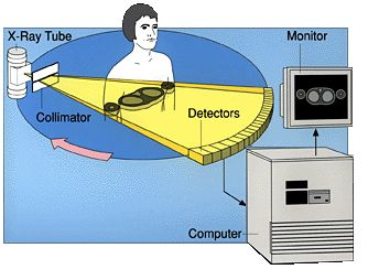
CT scanner, the machine used to proceed with computed
tomography, is composed of a rotating frame which has an x-ray tube on
one side
and a detector on the opposite side. As the rotating frame spins around
the
patient, a fan beam of x-ray continuously passes through the patient.
At each
rotation of the frame, the data stream representing the varying
radiographic
intensity sensed reaching the detectors is computer processed to
calculate
cross-sectional estimations of the radiographic density. A process
called
windowing calculates the radiographic density, expressed in Hounsfield
units,
to make very detailed images in 256 shades of gray. The shades vary in
function
of the density, becoming more white when more dense.
Often,
to accentuate the difference of intensity on the pictures, contrast
material
such as intravenous iodinated contrast or a dilute
suspension of barium sulfate will be
administered to the patient. It helps getting clearer images and it
highlights
structures difficult to see otherwise, like blood vessels. It
also helps to obtain functional
information about tissues.
Three dimensional reconstruction
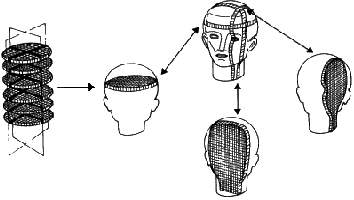

Another
method called volume rendering is also
commonly used to create three dimensional representations of the body
parts.
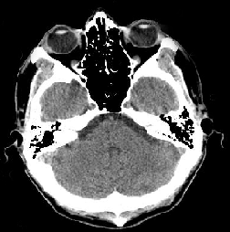
The image below shows several cross-sectional slices of head.
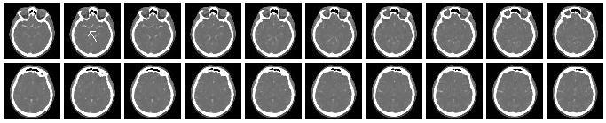
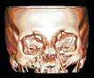
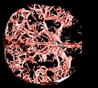
Using the same cross-sectional slices of head, it is possible to create a 3D representation of the internal structure of the head by a procedure called segmentation. It removes all the unwanted structures from the image and enables us to model, for example, the brain vessels.
Newer
machines with more advanced computer systems and
software strategies can process what is called helical or spiral CT.
Instead of
creating individual cross sections, the CT scanner processes
continuously
changing cross sections as the gantry with the patient is slowly slid
through
the X-ray circle. This procedure is very useful because it makes three
dimensional structures easier to create. Also, it may detect small
abnormal
areas better than do conventional CT. Moreover, it is faster, so the
test lasts
less long than with a conventional CT.
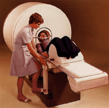
History
The first CT system was invented by British engineer Godfrey Newbold Hounsfield and by South Africa-born physicist Allan Cormack in 1972. The original prototype took several hours to acquire the data for a single semi-rotation, which took 160 parallel readings through 180 angles of 1° apart. The images were low resolution: about 80 x 80 pixels. Since that time, the speed and the resolution have considerably increased: today’s CT scanner reconstructs high resolution images from millions of data points in less than a second.
Uses
CT is used in the diagnosis of a large number of different disease entities and to determinate stage of cancer and to follow progress. But it is not yet well-performing in detection of tumors, coming behind magnetic resonance imaging.
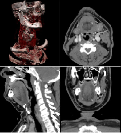
Cranial CT
Cranial
CT are most
frequently operated for the diagnosis of cerebrovascular accidents and
intracranial hemorrhage, but it is also useful to detect intracranial
pressure
and to evaluate facial and skull fractures. It is helpful for planning
craniofacial
and dentofacial deformities and to diagnose the causes of chronic
sinusitis.
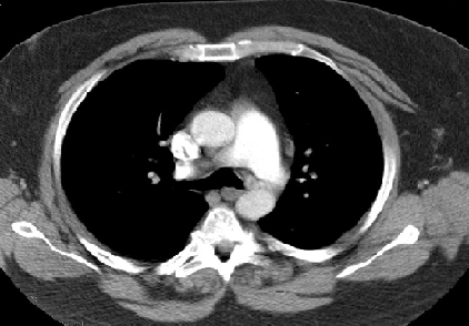
Chest CT
Different techniques of CT are used to detect and evaluate diseases related to the chest. CT is very useful to detect both acute and chronic changes in the lung parenchyma and to evaluate chronic interstitial processes such as emphysema and fibrosis. CT angiography of the chest is becoming the most efficient method to detect pulmonary embolism and aortic dissection.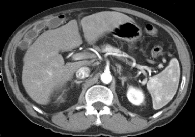
“CT is often used to image complex fractures, especially ones around joints, because of its ability to reconstruct the area of interest in multiple planes.”
Advantages
-A diagnosis effectuated with CT may eliminate the need for exploratory surgery.
-CT is completely painless.
-In comparison to conventional x-rays, CT scanning provides very detailed pictures of a large quantity of organs and tissues.
-CT can help planning radiotherapy
-The difference between tissues that differ in physical density by less than 1% can be distinguished because of the high contrast resolution.
-CT can be used for all anatomical regions, including those susceptible of patient motion or breathing because of the very short scan times varying from 500 milliseconds to a few seconds.
-Because of the possibility to create three-dimensional display, CT imaging is valuable for surgeonsTo obtain higher-resolution imaging and because of the complex scan techniques, the level of radiation varies from moderate to high. For example, in the most recent survey in the UK, CT scans constituted 7% of all radiologic examinations, but contributed to 47% of the total dose of x-rays emitted in medical examinations.
In
very rare cases, contrast agents may cause allergic symptoms such as
mild
itching or hives. Still in very rare cases, it can cause severe
allergic
reactions which include shortness of breath and swelling of the throat
or other
parts of the body. They can also induce kidney damage, principally to
patients
who have preexisting renal insufficiency, diabetes or reduced
intravascular
volume. For people who have a normal kidney function, the risks are
negligible
Computed
tomography is very useful to detect, diagnose
or evaluate physical troubles. Even if there are some risks related to
the use
of moderate to high x-rays doses, the considerable benefits of CT make
it a
method of medical imaging frequently used throughout the world
Sources
http://en.wikipedia.org/wiki/Computed_axial_tomography
http://www.chl.lu/html/glossaire_biomedical/computed_tomography.html
http://www.imaginis.com/ct-scan/
http://www.cancer.gov/cancertopics/factsheet/Detection/CT